
Blog
Share on social media
Teamwork makes the dream work - How a co-research approach tackles MRI challenges with AI
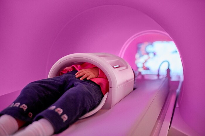
If you ever had an MRI yourself, you might have encountered an unpleasant experience. The MRI scan could be a lengthy procedure: the total examination can take from around 15 minutes up to one hour. During this time patients -often in pain- need to lay completely still, surrounded by noise in a confined space. This can lead to anxiety and claustrophobia and in the worst case, causes situations in which the scan needs to be repeated all over again. Furthermore, patients who move, result in suboptimal image quality leading to difficulties in reading the images by radiologist (diagnostic confidence) and maybe even repeating the entire examination. A new scan leads to stress for patients, increases in costs and additional pressure on the already tight schedule of the MRI-staff.
When we are able to minimalize the examination time, the added value for both patients and staff could increase.
So, you can imagine that when we are able to minimalize the examination time, the added value for both patients and staff could increase, not to mention the money that could be spend better. As our colleague – Richard Kemkers, Innovation Program Manager Precision Diagnosis - wrote in his blog about ‘the feel-good factor: why experiences matter to your radiology business’: we need to do a better job when it comes to how people feel about their care experiences.

AI-powered innovations: a step-change in MRI
Artificial Intelligence (AI) brings promising new possibilities to MRI scanning, as it can be applied to many aspects of the overall examination, like automated planning, segmentation and report creation. Scan acceleration is one of these new possibilities, as AI enables the acquisition of less data during an MRI examination, helping to reduce the time that a patient needs to be in the scanner. And that is exactly what we are aiming for when we combine accelerated scanning technology - Compressed SENSE - with AI-powered innovations: a step-change in MRI.
Artificial Intelligence brings promising new possibilities to MRI scanning.
Technologists are able to spend the gained time for instance to take more time for patient preparation and dealing with safety aspects. Moreover, by reducing the examination time, the patient’s experience improves and it makes MRI exams accessible for more people, like people who are in pain and who cannot lay still for a long period. All this off course helps improving the experience for patients and staff, enhance diagnostic confidence and increase productivity.
The FastMRI challenge
That is the reason why we were so proud and excited to be on stage December 2019 at the leading AI conference, NeurIPS in Vancouver in Canada. A panel of radiologists chose our team, with members from both Philips and our partner clinical partner at the Leiden University Medical Center (LUMC), as the top performer in the fastMRI image reconstruction challenge in the most clinically relevant challenge tracks. A second team in which Philips participated, together with the University of Amsterdam (UvA), the Amsterdam University Medical Center (Amsterdam UMC), and Radboud University, was the top performer in a separate category. The fastMRI challenge is a collaborative research project between Facebook AI Research (FAIR) and NYU Langone Health where AI and MRI specialists are being challenged to speed up MRI scans. The aim is to investigate the use of AI to make MRI scans up to 10 times faster. Thirty-four teams participated from around the world, using a number of AI solutions such as UNets, deformable convolutional networks, recurrent neural networks and other model architectures. All teams received the largest publicly available data set of de-identified raw MRI knee measurements that participants could use to train an algorithm that was then used to reconstruct hundred accelerated MRI scans. The winner came out after the top five entrants with the highest scores on a numerical metric, and later visually assessed by seven expert subspecialized musculoskeletal radiologists on several measures: contrast to noise ratio, artifacts, sharpness, diagnostic confidence, and overall image quality. The radiologists stressed the importance to visually asses the quality of the image, and to not rely blindly on numerical scores.
Quadruple Aim
With the quadruple aim we create solutions for value-based healthcare by improving patient and staff experience, with better outcomes and lower costs as a result. Improved Patient experience In radiology, patient anxiety over imaging procedures may impact the accuracy of diagnostic exams. The apprehension a patient experiences prior to an imaging study can impact results, skewing or delaying a diagnosis. In a study evaluating 172 patients undergoing diagnostic exams, 69% experienced high levels of anxiety, which can lead to hyperactivity of the autonomic nervous system and produce symptoms that can directly influence exam results. In another study, anxiety reactions – including increased heart rate and blood pressure – were reported in up to 30% of patients undergoing MRI scans. Improved Staff experience Most of the time MRI-staff work in a tight and challenging schedule. The increasing pressure to realize lower healthcare costs, combined with the growing demand for medical imaging results in a higher workload. This makes it more likely that they will experience burnout. This is illustrated by recent research (2019) we conducted in France, the US, UK and Germany showed alarming numbers of technologists report moderate or severe levels of job stress (FR=40%; US=44%; UK=54%; GER=97%). [3] Technologists in these countries also reported a significant incidence of moderate or high burnout (UK=30%; FR=33%; US=36%; GER=97%). Taken together with burnout levels for radiologists, we’re witnessing a serious, systemic problem across imaging. Better Outcomes The radiology department plays a critical role in diagnosing and guiding patients into the right treatment plans. Today, advances in medical imaging technology and radiology protocols hold tremendous potential to help radiologists reach more consistent, confident diagnoses faster. [4] Lower cost of care Over the past few years MRI has experienced tremendous growth in the number of scans performed annually, due in large part to an increase in chronic diseases and a growing aging population. At the same time, MRI scanners are becoming faster with techniques like Compressed SENSE. These technology advancements not only contribute to faster turnaround times and a better patient experience, but help boost overall productivity of imaging departments.

Teamwork makes the dream work
Winning the challenge was not the only reason for our excitement, the process towards this great achievement made us also extremely proud. Key to our success was a multi-disciplinary team involving experts and technicians in the field of AI and MRI and clinical partners. The team included experts from both Philips and LUMC working fulltime for months on the AI algorithm, leading to this great success.
Key to our success was a multi-disciplinary team involving experts and technicians in the field of AI and MRI and clinical partners.
The close and intense partnership we had with LUMC during this challenge was defined by our mutual target, focus and clear deadline. Our partnership with LUMC kicked off in June, with extra people in the team to help cracking the nut. We adopted an Agile way of working, with daily conversations and weekly assessments of the research output and parallel research tracks, allowing to explore the most promising ideas more in detail. Our key learning was that you really move forward if you receive feedback from different related environments. Of particular importance was the involvement of radiologists and clinical scientists who assessed the quality of the reconstructed images from a clinical point of view. A partnership like this requires more than investing in terms of money and time, it relies on knowledge sharing, and for that you need mutual trust.
A partnership like this requires more than investing in terms of money and time, it relies on knowledge sharing, and for that you need mutual trust.
The team demonstrated that - in little over 6 months – it is possible to deliver breakthrough solutions via excellent teamwork. The cooperative working atmosphere and the mutual trust that we now share will definitely pave the way for other innovative projects, including the clear focus in bringing this success to the patients as soon as possible. [1] F. Muscarneri. Evaluation of anxiety level in patients waiting to undergo diagnostic radiological exams. European Society of Radiology. 2013 [2] Grey SJ, Price G, Mathews A. Reduction of anxiety during MR imaging: a controlled trial. Magn Reson Imaging. 2000; 18:351-55 [4] https://www.usa.philips.com/healthcare/medical-specialties/radiology
Nicola Pezzotti Research Scientist in Artificial Intelligence Nicola Pezzotti joined Philips Research in 2018, where he researches AI techniques for medical imaging and keeps close relationships with academic partners. Besides his experience in the startup world, he obtained the PhD Cum Laude from Delft University of Technology and joined INRIA and Google AI as visiting scientist. He is recipient of several awards, including the IEEE VGTC Best Dissertation Award and the Dirk Bartz Prize for visual computing in medicine.
Elwin de Weerdt Reconstruction Algorithm Architect Philips MR Elwin is passionate about developing breakthrough, meaningful innovations. In his role he is responsible for translating and shaping innovations along the path from research to product. One of the latest innovations he worked on as Technical Lead was Compressed SENSE, a breakthrough innovation in MR scan acceleration.