In order to make a definitive diagnosis it is then necessary to take a biopsy, following which a pathology test is carried out. It is not easy to take a good biopsy, and ‘false positive’ results are therefore not uncommon. One solution we have recently brought to market is a technology that uses existing MRI images in combination with ultrasound while the biopsy is in progress. This makes it possible to visualize the prostate and the tumor within it. A small electromagnet at the tip of the biopsy needle allows it to be guided with precision to the tumor tissue that can be seen in the merged MRI/ultrasound images.
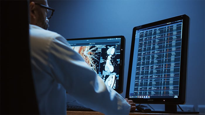
Digital technologies have recently also made it possible to automate the way in which pathology tests are carried out – until today these tests have not been automated at all. Digital pathology makes it possible to record microscopic tissue images, to share them and even to obtain a digital second opinion.
If we look at genomics and the invention of targeted drugs, we can see that next-generation sequencing, or the unraveling of the entire DNA pattern or RNA pattern, for instance in cancerous tissue, is within reach. In combination with bio-informatics, we are already able to obtain some indication beforehand as to whether or not a therapy will work for a given patient because we are able to detect the activated biomolecular cancer mechanism.
In order to make a definitive diagnosis, it is essential that all the available information is brought together. Philips is currently developing software for an interdisciplinary tumor board, a kind of cockpit that can bring together MRI images, the images from digital pathology and genetic information, among other things, so that it becomes much easier to discuss, share and record outcomes. This kind of portal or app can be helpful for patients as well.
By providing specific and personal information about the diagnosis and the patients themselves – at their level of knowledge – it is possible for patients to read about the pros and cons of the various treatment options and to have them explained to them in their own time. This also gives them a chance to discuss the situation and the relevant information with their family and friends at a time that suits them. That is important because there is often a whole plethora of (surgical) treatment options and medicines from which the patient can choose. And in some cases it can even be better to do nothing at all for the time being; the ‘watchful waiting’ approach is sometimes adopted for mild forms of prostate cancer, for example.
Treatment: Less Invasive Forms of Therapy
New developments make it possible to use less invasive forms of treatment. One example is ablation. This involves using high-intensity focused ultrasound to heat the tumor tissue to such an extent that the proteins become denatured and die. MR images can be taken while this is being done, thus making it possible to very accurately target the area to be treated in the tumor tissue.
Another minimally invasive treatment method that has become available on the market is image-guided trans-arterial chemio-embolization. This method uses the existing catheter lab set-up that is commonly used for endovascular treatments. Tumors attach themselves to the regular blood flow with their own feeding vessels, in order to grow faster. Embolization is a technique where an image-guided catheter is inserted into these newly created blood vessels so that a highly concentrated chemo agent can be applied loco-regionally. The treated region is subsequently sealed via embolization to terminate the blood supply.
A very exciting new technology for treating tumors is image-guided radiation therapy. Philips is working together with Elekta and the University of Utrecht on the MR-LINAC – in which extremely precise MR imaging can be used to continuously adjust the radiation beam to compensate for any movement of, for instance, a tumor due to the patient breathing. It is hoped that this will ultimately help to reduce the damaging side effects of the treatment.