Innovation Spotlight: Finding new ways of enhancing magnetic resonance imaging
Apr 15, 2025 | 5 minute read
About the author About the author
A team of writers, editors and visual designers covering the latest trends and developments in healthcare, focusing on the role of innovation, design and sustainability in improving health outcomes.
You are about to visit a Philips global content page
Continue In 1973, Nottingham University physicist, Sir Peter Mansfield, first harnessed nuclear magnetic resonance to create cross-sectional images of living tissue. This was the beginning of a revolution in medical diagnosis: magnetic resonance imaging (MRI).
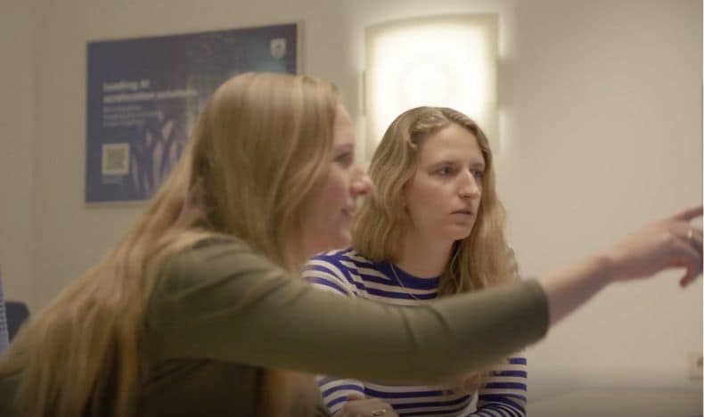
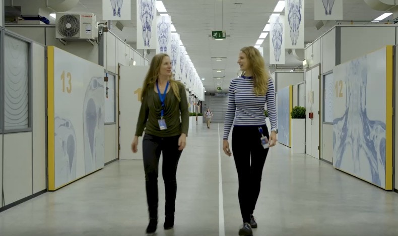
Now, with over 60 million scans conducted in hospitals around the world each year, MRI technology remains the gold standard for soft tissue imaging, used extensively for diagnosing and treating various health conditions. At Philips, we have been continuously innovating this life-saving technology, finding new ways to enhance imaging through artificial intelligence (AI) enablement and leveraging data use. At the forefront of MRI innovations in Philips are Yolanda Noorda and Kirsten Koolstra, MRI Reconstruction Algorithm Developers, transforming data into images through algorithms that enhance medical imaging to speed up time to diagnosis. Let’s find out about their incredible work and the role they’ve played in helping to revolutionize MRI technology.
Tell us about your roles at Philips: What led you here, and what brings you to work every day?
Yolanda: I’m the Group Leader of the MRI Reconstruction team in Best and have been a member of this team for more than eight years. I knew I wanted to join Philips as soon as I went into healthcare; in the Netherlands, it’s the place to go if you want to make an impact on the world and improve MRI. Developing algorithms that make a difference in people’s lives is very rewarding. It takes a lot to bring a product from concept to market, and it's incredibly fulfilling to overcome those challenges. You work together with lots of different people with their own expertise and background, so you learn a lot. Kirsten: I’m an MRI Reconstruction Algorithm Developer and I’ve been here for almost three years. I love working on solutions that help people reach a precise diagnosis the first time. I really like the problem-solving aspect: there are always moments when your results don't match your expectations, and you need to dig deeper to find out why. I joined Philips after working in MRI research, and I wanted to see how it translates into becoming a product. I struggled with making the jump from academia to industry for a long time, and finally recognized that I’d never know what it's like unless I tried it. I’m really happy that I got the chance to take this step. Working on MRI reconstruction in a Dutch company is quite unique.
Tell us about the work you do in MRI reconstruction. What is it, and how has AI changed the game?
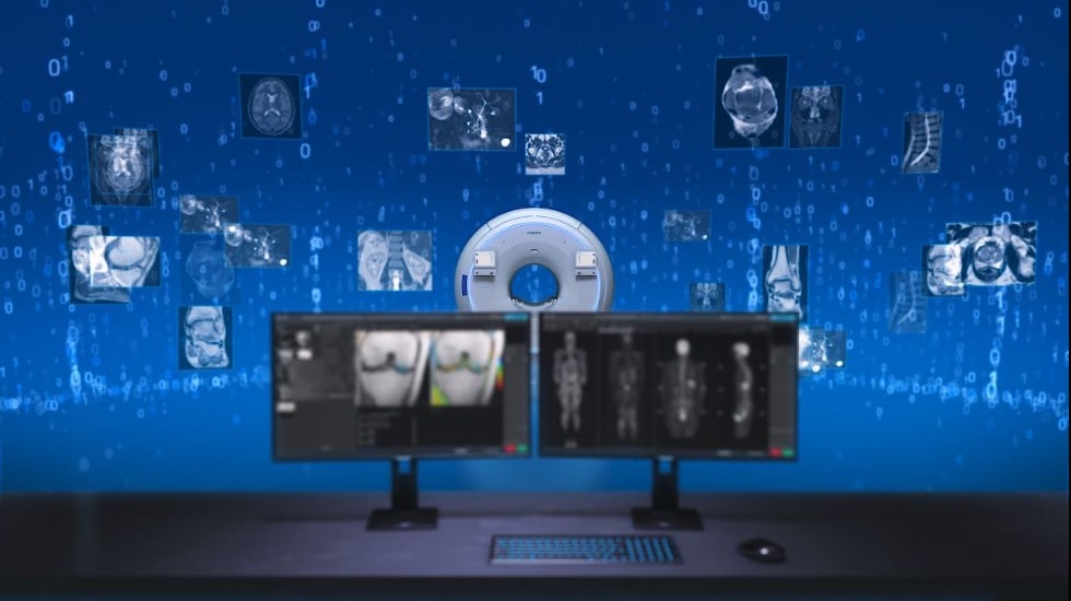
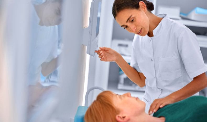
Kirsten: When a patient is lying in the scanner, the magnetic coils that surround them generate a signal, which gives us the information we need to reconstruct a diagnostic image. The problem is, MRI generates a lot of noise and artifacts, too. Our task is to “denoise” MRI images: i.e., filter noise out to only focus on the clinically useful data underneath so that we get accurate, high-quality images – quickly.
Yolanda: For example, our MRI SmartSpeed software uses Adaptive-CS-Net, an AI reconstruction algorithm to accelerate MRI scans. By applying AI algorithms during image reconstruction, it achieves up to 65% greater resolution* and up to 50% shorter exam times†. SmartSpeed also enables us to leverage all the scan data to denoise the image as soon as possible in the reconstruction process, creating a better-quality image faster than before – going beyond what classical reconstruction techniques can do.
Kirsten: We train our algorithms to split the noise from the signal by detecting which is which. We apply this learning to newly seen scan data, improving image quality and decreasing scan time. However, developing an algorithm is just the start of our product development. To get it ready for market, we need to standardize it for usefulness across different clinical applications, ensure stability and safety through rigorous testing, and design the product to optimize user-friendliness.
Why is reducing scan time so important?
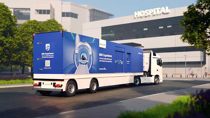
Yolanda: MRI is a slow process, so innovation that speeds it up is crucial. Some complex scans can easily take up to 45 minutes. Reducing this means patients spend less time laying still in the scanner, and more patients can have access to the technology. Just recently, we showcased the latest release on the software, SmartSpeed Precise, at the European Congress of Radiology (ECR). This development further reduces scan times and improves diagnostic image quality, extending AI-driven efficiency across the entire Philips MR portfolio.
How does your work contribute to solving current challenges in clinical practice?
Kirsten: Before I came to Philips, I worked in a hospital and saw firsthand the pressure of waitlists and long hours. It's exciting to know my work can help alleviate that pressure for both patients and staff. Yolanda: We’re close to our customers, so we have a great idea of what they need from our products. Philips regularly visits hospitals to observe how clinicians really work with our products so we can refine them even further using those insights.
What do you see in the future of MRI?
Yolanda: At Philips, we are dedicated to the ongoing transformation of healthcare delivery by integrating informatics and AI and, last year, we were the leading applicant in the field of medical technology at the European Patent Office. Our team will continue to contribute to driving innovation and, in the future, as scanning becomes more automated and efficient, we’ll be able to further increase first time right scanning and shorten total exam times. In turn, this will continue to decrease the need for rescans and enable patients to get diagnosed quicker. AI could be involved in every aspect – not just in reconstruction but in creating smoother, patient-specific workflows.
*Compared to Philips SENSE imaging. † Compared to Philips scans without Compressed SENSE. “The opinions and clinical experiences presented herein are specific to the featured article and are for information purposes only. The results from and experiences may not be predictive of all patients. Individual results may vary depending on a variety of patient-specific attributes and related factors. Nothing in this presentation is intended to provide specific medical advice or to take the place of written law or regulations.”