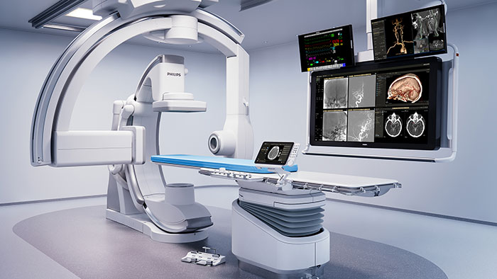
IVUS and iFR technologies receive top-level recognition in new ESC Guidelines, reinforcing their role in optimizing coronary interventions and patient outcomes
Sep 30, 2024 | 4 minute read
Amsterdam, the Netherlands – Royal Philips (NYSE: PHG, AEX: PHIA), a global leader in health technology, acknowledged today the latest classification updates for the company’s key imaging and physiology technology in new guidelines for the management of chronic coronary syndromes from the European Society of Cardiology (ESC).

The latest updates to these ESC guidelines emphasize the robust clinical evidence supporting the use of intravascular ultrasound (IVUS) and instantaneous wave-free ratio (iFR), both of which have demonstrated significant benefits in improving patient outcomes through numerous large-scale randomized trials like DEFINE FLAIR and iFR SWEDEHEART. The updates were announced at the recent ESC Congress in London, five years after the last update in 2019, and simultaneously published in the European Heart Journal.
In the 2024 ESC Guidelines for management of chronic coronary syndromes, several Philips technologies for coronary artery disease (CAD) were included, reflecting their level of evidence and the evolution of clinical science establishing standards of care to optimize patient outcomes. Class guidelines denote the evidence a treatment or procedure is recommended or indicated for use, whereas the level of evidence is based on the amount of data available from randomized clinical trials and meta-analyses.
IVUS for intracoronary imaging upgraded to Class IA recommendation
“Meta-analysis of randomized clinical trials”—Stone et. al [3]—“had already shown that intracoronary image guidance of PCI improves patient outcomes and saves lives,” said Javier Escaned, MD, Head of the Interventional Cardiology Section at Hospital Clinico San Carlos, Madrid Spain. “But the IA recommendation for IVUS in the updated ESC guidelines is crucial, as it reflects expert consensus based on a definite body of evidence supporting the positive impact of IVUS, specifically for patients with anatomically complex lesions treated with PCI.”
But the IA recommendation for IVUS in the updated ESC guidelines is crucial, as it reflects expert consensus based on a definite body of evidence supporting the positive impact of IVUS, specifically for patients with anatomically complex lesions treated with PCI.
Philips intravascular ultrasound (IVUS) technology is an innovative imaging tool used during coronary interventions that provides detailed, real-time cross-sectional images of the inside of blood vessels, allowing for precise assessment of vessel anatomy and plaque characteristics to optimize stent placement to improve outcomes. This technology has been upgraded to Class 1, Level of Evidence A, reflecting the growing body of evidence showing that intravascular imaging (IVI) improves patient outcomes. With the updated guidelines, IVUS is now recommended for percutaneous coronary intervention (PCI) on anatomically complex lesions, in particular left main stem [4], true bifurcations [5] and long lesions [6].
“The percentage of PCI considered to be complex is increasing, with current estimates around 40-50% or more [7] The increasing complexity of these interventions underscores the need for intracoronary imaging to be used much more frequently than it is today,” said Stacy Beske, Business Leader for Image Guided Therapy Devices at Philips. “Philips plug-and-play digital IVUS on IntraSight offers an easy and seamless way to bring the benefits of intracoronary imaging to patients and clinicians.”
iFR and FFR for physiologic measurement during PCI remains Class IA recommendation
A second Philips technology that was included in the new ESC Guidelines is Philips’ instantaneous wave-free ratio (iFR), a physiological measurement used during percutaneous coronary intervention (PCI) to assess the severity of a coronary artery stenosis without the need for inducing hyperemia, enabling real-time decision-making about the necessity of stenting. Philips iFR remains the gold standard for physiologic measurement during PCI [2] and is the only resting index called out as Class I recommendation, level of evidence A, alongside fractional flow reserve (FFR) [1,10].
This classification can be attributed to two of the largest physiological studies completed, DEFINE FLAIR and iFR SWEDEHEART, which involved more than 4,500 patients combined and evaluated the safety and effectiveness of iFR, demonstrating its non-inferiority to fractional flow reserve (FFR) [8, 9].
For patients with non-obstructive CAD, coronary function testing, such as coronary flow reserve, has been elevated to Class 1, Level of Evidence B. This technology addresses patients with non-obstructive CAD by identifying potentially treatable endotypes and offering a definitive diagnosis of microvascular disease that can direct medical treatment strategies. This change to the guidelines validates the growing prevalence and diagnosis of microvascular disease.
Philips CAD solutions consist of advanced clinical and workflow applications, therapeutic and diagnostic devices, and leading services, which work together to efficiently support every step of coronary procedures. From diagnosis to restoring vessel patency, the coronary suite ecosystem is designed to enhance performance and efficiency.
Sources [1] 2024 ESC Guidelines for the management of chronic coronary syndromes. (European Heart Journal; 2024 – doi: 10.1093/eurheartj/ehae177)
[2] Lawton J. et al. 2021 ACC/AHA/SCAI Guideline for Coronary Artery Revascularization. JACC. 2022;79(2):e21-e129.
[3] Stone G, et al. Intravascular imaging-guided coronary drug-eluting stent implantation: an updated network meta-analysis. The Lancet, Volume 403, Issue 10429, 824 - 837
[4] The left main stem (LMS), also known as the left main coronary artery (LMCA), is a critically important artery that serves as the primary blood supply to a large portion of the heart
[5] True refer to a type of coronary artery bifurcation lesion where there is significant disease (stenosis or narrowing) involving both the main branch (main vessel) and the side branch (branching vessel) at the point where they diverge from each other
[6] Coronary artery blockages that extend over a longer segment of the artery
[7] Hanna, Wang, Kochar, et al. Complex Percutaneous Coronary Intervention Outcomes in Older Adults JAHA 2023; 12; 19.
[8] Gotberg M., Christiansen E.H., Gudmundsdottir I.J. Instantaneous wave-free ratio versus fractional flow reserve to guide PCI. N Engl J Med. 2017;376:1813–1823.
[9] Davies J.E., Sen S., Dehbi H.-M. Use of the instantaneous wave-free ratio or fractional flow reserve in PCI. N Engl J Med. 2017;376:1824–1834.
[10] Lawton J. et al. 2021 ACC/AHA/SCAI Guideline for Coronary Artery Revascularization. JACC. 2022;79(2):e21-e129.


