Philips advances minimally invasive therapy procedures in prostate cancer care with FDA 510(k) clearance for image-guided navigation technology
Supporting prostate cancer diagnosis with more precise guidance for clinicians, offering better patient care
Jul 23, 2025 | 3 minute read
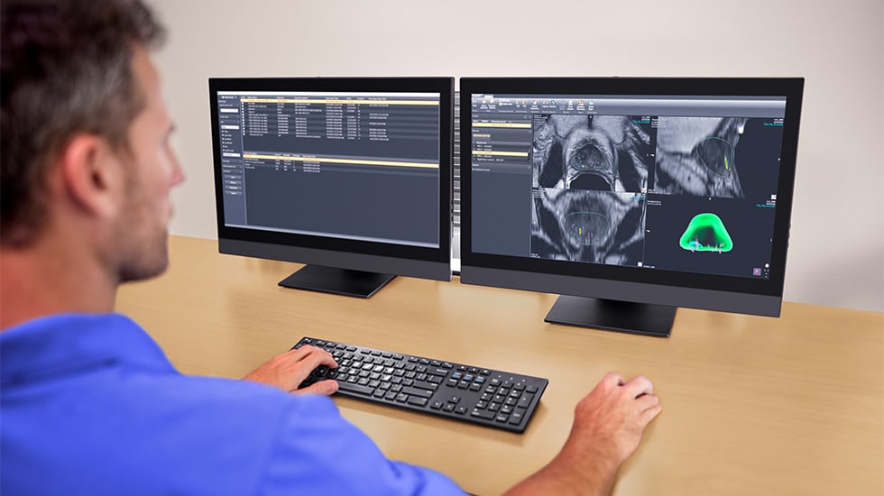
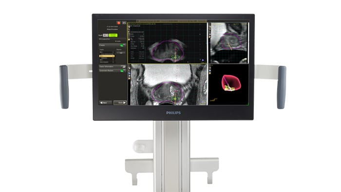
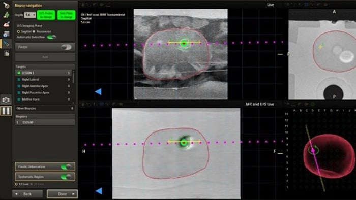
Amsterdam, the Netherlands – Royal Philips (NYSE: PHG, AEX: PHIA), a global leader in health technology, today announced a major advancement in image-guided navigation for prostate cancer care with the FDA 510(k) clearance of the latest Philips UroNav version. The system includes a new advanced annotation [1] workflow that supports clinicians during focal therapy procedures, helping deliver more precise, minimally invasive care.
This release comes at a time of increasing need for innovation in prostate cancer care. Prostate cancer remains the most commonly diagnosed solid tumor among men in the United States, with over 313,000 new cases, 1 in 8 men will be diagnosed with prostate cancer in their lifetime [2]. While many prostate cancers are slow-growing, overtreatment can lead to quality-of-life degradation, including incontinence and sexual dysfunction, prompting growing interest in focal therapies that offer a targeted, tissue-sparing alternative for appropriate patients.
With fused imaging and real time ablation guidance in one place, we can personalize therapy with greater accuracy and spare patients the unnecessary side effects of traditional treatments.
“We’re entering a new era of precision prostate cancer care. Philips’ integrated focal therapy platform unifies imaging, biopsy pathology, treatment planning and 3D imaging guidance with MR US fusion giving clinicians end to end efficiency and control,” said Dr. Ardeshir Rastinehad, Vice Chair of Urology at Lenox Hill Hospital and System Director of Prostate Cancer at Northwell Health. “With fused imaging and real time ablation guidance in one place, we can personalize therapy with greater accuracy and spare patients the unnecessary side effects of traditional treatments.”
As an image fusion system, UroNav seamlessly integrates pre-procedural imaging, such as Magnetic Resonance Imaging (MRI), with real-time intra-procedural imaging from ultrasound (US) systems. This innovative combination enhances the precision and accuracy of therapeutic procedures, providing clinicians with a comprehensive and dynamic view of the targeted area.
A more targeted approach means a more informed treatment selection and patients receive better and more precise care and clinicians are supported in better diagnosis, a 30% improvement in high-risk prostate cancer diagnosis using fusion biopsy compared to the standard biopsy [3]. The new advanced annotation workflow works in tandem with DynaCAD Urology to support focal therapy planning, deliver and review, reducing complexity and enabling a broader group of clinicians to offer minimally invasive options.
“We’re helping clinicians deliver more precise prostate cancer care by streamlining complex workflows and delivering the insights they need to support precise diagnosis and expand options for minimally invasive treatments,” said Martijn Hartjes, Business Leader, Clinical Informatics at Philips. “Our goal is to equip clinicians with the clinical tools required so they can deliver better care for more patients.”
In addition to clinical functionality, Philips UroNav delivers enhanced compatibility with ultrasound devices and needle guides, upgraded privacy and security protections, and seamless integration with Philips DynaCAD systems for radiology and urology.
Philips’ comprehensive urology portfolio integrates imaging, biopsy, therapy guidance, and digital pathology to support precision diagnosis and treatment—enabling clinicians to tailor care with greater efficiency and confidence.
Sources [1] The advanced annotation option is sold separately. The software is not intended for diagnosis and is not intended to predict ablation volumes or predict ablation success.
[2] Key Statistics for Prostate Cancer. American Cancer Society. https://www.cancer.org/cancer/types/prostate-cancer/about/key-statistics.html
[3] Siddiqui MM, et al. Comparison of MR/ultrasound fusion–guided biopsy with ultrasound-guided biopsy for the diagnosis of prostate cancer. JAMA. 2015;313(4):390-397.

Media contacts

Contact details Contact details
You are about to visit a Philips global content page
Continue