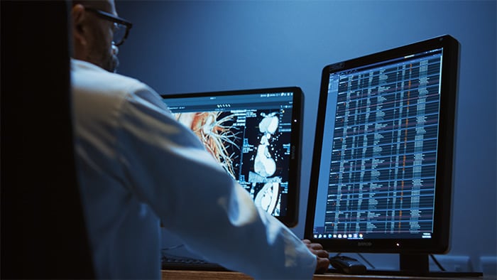
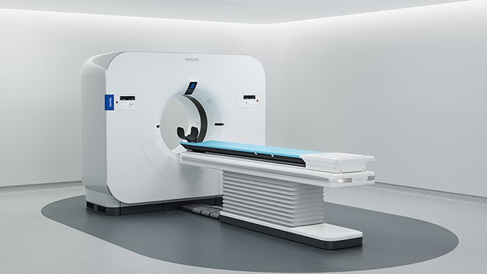
Yet, according to a 2016 TMTG study, 70% of radiologists still consider neuro indications to be challenging, mostly due to a lack of imaging and visualization techniques. We believe that magnetic resonance imaging is in a unique position to address neurological disorders and has the potential to bring diagnostic clarity, even to difficult neurological cases. Today’s MR tools can shed light on intricate structures in the brain, providing vital insights. And advanced visualization applications help clinicians review complex, multi-dimensional data to make informed, definitive decisions. Elevating neuro diagnostics So what is driving the need for greater insight into the brain? Around the globe, the burden of neurological disease is rising. Demographic changes such as aging populations in many parts of the world are mostly behind these developments. And, naturally, healthcare providers need to respond. But do they have the tools they need? Philips is committed to pushing the limits of neurological imaging and elevating neuro diagnostics. This year, we are introducing a set of novel imaging and visualization strategies that leverage our dStream digital platform. These may empower you to resolve complex neuro questions with more certainty, as well as unlock new territories in advanced neurofunctional applications. With this enhanced portfolio of applications, Philips aims to provide the best possible diagnostic clarity and treatment guidance for all patients with neurological disorders. Moreover, it is a key step on our journey to elevate neuro diagnostics, make MR more definitive[1], and touch more lives with this advanced technology. Learn more about MR neuro imaging here. Enhancing diagnostic confidence for brain imaging Philips MR neuro applications let you harness the power of MR to address challenges for example in brain tumors and vascular diseases. Black Blood imaging, for example, helps you better differentiate the vessel lumen from the intra lumen blood signal. This enhances diagnostic confidence by letting you perform 3D brain imaging with higher isotropic resolution[2] with a reduction of the intra-lumen brain blood signal[3] over the complete imaging volume. Plus, 3D isotropic acquisition enables reformats in any plane (including oblique) without loss of resolution. What’s more, another key advantage is that you can perform scans in just five minutes[4].
Black Blood imaging helps you get a clear view of vessel lumen A personalized approach As I already mentioned, recent years have seen a sharp rise in the cases of neurological disorders. But it’s easy to forget that we’re not simply talking about statistics here: behind each and every one of these diagnoses is a patient. And each one of them has their own expectations, needs and story. Tailoring healthcare to meet individual requirements is a key challenge today. It is also something that is very close to our hearts. That is why we intend to continue to push the envelope in neuro imaging, e.g. towards quantitative biomarkers to support personalized diagnosis and treatment guidance. This approach not only paves the way for evidence based outcomes, but it also helps you deliver care that is in line with your patients’ unique requirements. Embracing the future As we have seen, methods for studying the brain and diagnosing neurological conditions have come a long, long way. As society changes, MR techniques and solutions have evolved to meet new requirements. But it doesn’t stop here. It’s an ongoing journey – and one that we are incredibly proud to be on. It may sound clichéd to say “the future starts now”, but the new neuro applications we’re unveiling at RSNA can already provide a glimpse of where the MR neuro journey may lead. To learn more about this topic and see what else we’re doing this year, check out our website or watch this video. [1] Definitive is defined as features that are expected to deliver alternative contrasts, functional or quantitative images. [2] Compared to Philips 2D double inversion methods with same brain coverage and scan time. [3] Compared to Philips 3D T1w scan without MSDE pre-pulse. [4] Compared to Philips 2D double inversion recovery methods with same full brain coverage.
You are about to visit a Philips global content page
ContinueYou are about to visit the Philips USA website.
I understand