By Dr. Junzi Shi, MD
Dr. Shi is a radiology resident at Brigham & Women’s Hospital in Boston, MA and a clinical fellow at Harvard Medical School. She has written articles on physician finance, patient safety, medical education, and improving the patient experience.
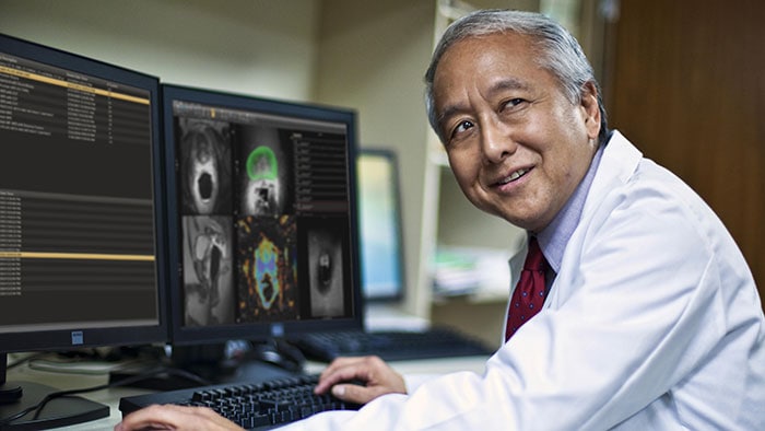
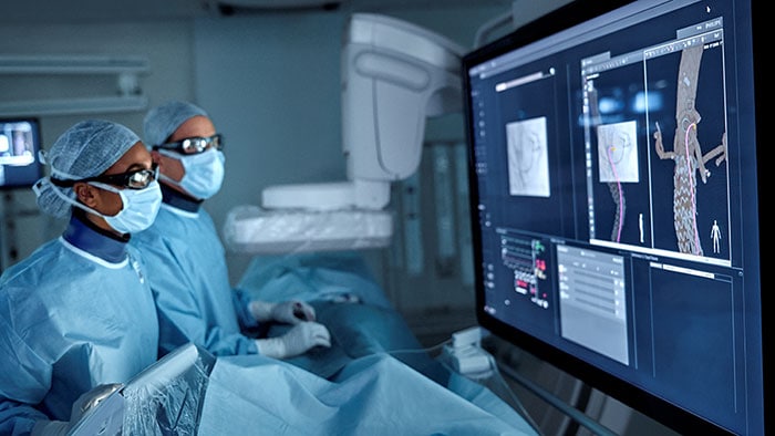
Over the past 50 years, the ability of imaging to detect and diagnose abnormalities in the human body has improved tremendously. Radiologists are the doctors responsible for interpreting these imaging tests and performing advanced imaging research within both diagnostic and interventional subspecialties. A diagnostic radiologist’s primary role is interpreting imaging exams within the reading room, while an interventional radiologist performs image-guided, minimally-invasive procedures in an operating room setting.
Radiologists work with physicians from different disciplines, technologists, nurses and other staff as an organized team. From this group, patients will tend to interact most with technologists, who guide them through the test.
Radiologists go through more medical training than many primary care physicians. In a recent survey on patient perception and understanding of radiologists, only 12% of respondents correctly identified that radiologists obtain 14 years of post-high school training. The lack of public understanding spurred the American College of Radiology to start the ‘Face of Radiology’ campaign to help increase awareness of the discipline and the fact that cutting-edge research led by radiologists has redefined and reshaped medicine.
While some referring physicians and specialists may look at the scans on their own, a formal radiology interpretation is required for each study. In their reports, radiologists describe findings and summarize key points in the ‘assessment and recommendation’ section. If there are concerning results, the radiologist will contact the referring physician and have a discussion about the next steps. In addition, radiologists are stewards of imaging resources and monitor radiation doses to minimize risk to the patient.
As a radiologist, I see a variety of image-related problems that occur during the exam.
As a radiologist, I see a variety of image-related problems that occur during the exam. The most common problem is an artifact – something observed in the image that is not naturally present but occurs as a result of the investigative procedure – that comes from body movement or breathing. Similar to someone moving while a photo of them is being taken, this causes blurring of the image and decreases the ability of the radiologist to make an accurate diagnosis.
Another important and common artifact seen on both CT and MRI exams is caused by metal on the body – such as dental hardware, jewelry or implants. Even small pieces of metal can cause large lines obscuring the image, making it important to remove all the metal possible before starting the test. Sometimes the artifact is unavoidable and radiologists simply have to do their best to ‘read through’ the obstruction.
Face-to-face radiology consultations are becoming increasingly popular. Surveys have shown that up to 90% of patients are interested in meeting with the radiologist to hear the results at the time of the procedure (Domina 2016, Rosenkrantz 2015, Mangano 2014), with 57% willing to pay for the service. Speaking with the radiologist directly may provide a more in-depth explanation than simply hearing the summary from another physician. Under the US healthcare system, taking time for patient consultations is not currently reimbursed for radiologists and remains a sticking point. If these patient interactions can be covered by insurance or paid out of pocket, this service may become more readily available.
Some centers have already taken the leap and implemented dedicated radiology clinics. For example, Cincinnati Children’s Medical Center offers a consultation service that allows radiologists to deliver results directly to patients after the completion of the test, helping to clarify details and answer any questions. This reduces anxiety for some patients, while others may prefer to obtain the results from their primary care physician with whom they have already established a relationship and treatment plan.
Radiology, though, is the future of medicine. The ability to ‘see inside the body’ using a variety of techniques is a privilege and highly sought-after skill. Improved CT and MRI scanners have already drastically increased the sensitivity and specificity of diagnostic scans and interventional radiology provides a variety of minimally invasive solutions. As we gain more knowledge about various disease processes, there will be a greater role for radiology in the prevention of disease.
Perhaps one day we will have a subspecialty of ‘preventative radiology’ that combines genomics and metabolism with imaging. The interaction of genotype and the environment results in observable characteristics, including what we can see with imaging. In the future we may keep ‘baseline’ whole body scans as part of each patient’s medical profile in order to provide individualized care. Baseline imaging can provide a valuable insight to the current state of our bodies, while whole genome sequencing may identify genes that are risk factors for various diseases.
This is not currently recommended due to the lack of a favorable cost-risk-benefit ratio, but further advances in knowledge and technology are rapidly coming down the path. The combination of genomics, whole body scans and functional testing may help further translate personal health data into estimated risk and, ultimately, improved healthcare. The possibilities are endless and the number of potential lives saved infinite.