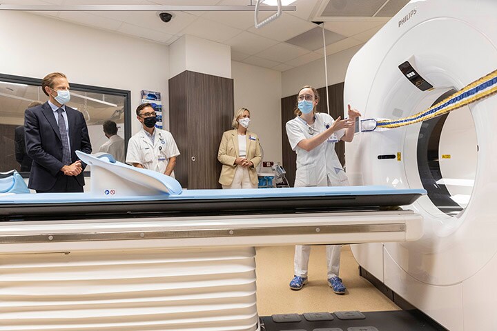
This month, University Medical Center Utrecht (UMC Utrecht) in the Netherlands officially opened its new ‘CT-Street’ - a suite of state-of-the-art spectral CT scanners from Philips that speed up CT scans while at the same time enhancing image quality and detail. UMC Utrecht is the first medical center in the world to be equipped with two of these advanced Philips imaging solutions.
The big advantage of these new systems is that we can get much more information from the scans using the same amount of radiation.
Marjan Hennipman
Unit Head of the Radiology Department at UMC Utrecht
"The big advantage of these new systems is that we can get much more information from the scans using the same amount of radiation,” Marjan Hennipman, Unit Head of the Radiology Department at UMC Utrecht. “In addition, scans need to be repeated less often, which is also a great advantage for patients." UMC Utrecht’s purchase of these new scanners is in line with its policy of using high quality imaging to provide patients with more targeted treatment. The Philips Spectral CT scanners, which incorporate a dual-layer spectral detector that differentiates between high energy and low energy X-ray photons to provide spectral images, enhance UMC Utrecht’s diagnostic capabilities so that its medical teams can quickly provide a reliable diagnosis and an effective treatment plan for the patient. In addition, the patient does not have to spend as long in the scanner, because the scans are completed within seconds. "With the help of spectral information, a diagnosis can be made more often in one go. For example, a blood clot can be found more easily, the blood flow in organs can be deduced, and information can also be obtained about the tissue composition of a possible tumor,” said Professor Pim de Jong, Medical Department Head of Radiology at UMC Utrecht. “In principle, a lot of additional information can be obtained about the general condition of a patient, such as bone density and muscle quality. Finally, we want to investigate whether this method is also more sustainable, for example, in terms of using less contrast agent."

The Philips CT systems will be used for complex imaging of many different internal organs, as well as for cancer imaging and image-guided treatment. The system helps to reduce the number of repeat and follow-up scans. In addition to using the company’s CT systems, UMC Utrecht and Philips have worked together in the fields of radiology and radiotherapy for many years to develop new techniques and innovations, including in MR imaging, with the aim of enhancing patient outcomes and patient and staff experiences, and reducing the cost of care. “For many years, we have been focusing on image guided therapy at UMC Utrecht”, said Anouk Vermeer, board member at the hospital. “We have spearheaded the development of minimally invasive treatments. About twenty-five years ago, our colleague Jan Lagendijk, professor of clinical physics, devised the MR-Linac, which provides a more accurate form of radiation treatment because the doctors can better see where the tumor is. Together with the business community, including Philips, this idea has been further developed and many new applications have been added.” "Together with UMC Utrecht, we are continuously working to improve patient care," said Henk Valk, CEO at Philips Benelux. "We not only provide the latest and most advanced technology, but also work together on the development of new treatment methods and technologies for diagnosis. UMC Utrecht is a world-leading academic center where together we conduct research that is at the core of global innovations in healthcare. I am very proud of the long-standing and successful cooperation we have with UMC Utrecht."
Share on social media
Topics
Contact

Kathy O'Reilly
Philips Global Press Office Tel.: +1 978-221-8919
You are about to visit a Philips global content page
Continue


