Enabling first-time-right imaging for every child – Q&A with Atul Gupta, MD
Jun 04, 2024 | 8 minute read
Diagnostic imaging has become a standard and indispensable element of almost every patient care pathway. But for pediatric patients – from newborns to teenagers – acquiring precise diagnostic images quickly and safely comes with unique challenges, requiring dedicated teams, procedures, interventions, and technology.
In pediatric care, time is survival. Disease can progress quickly in pediatric patients – for instance, in children with solid malignancies, we know that just a six-month delay in diagnosis results in a five-year survival decline: from 73% to 40%. [1] For this reason, achieving first-time-right appropriate and high-quality imaging is vital to getting accurate diagnoses, accelerating time to treatment, and achieving the best treatment outcomes. The challenges with pediatric imaging mostly boil down to motion, safety, and access to care. Fortunately, solutions are being developed to address many of these challenges which benefit both patient and caregiver. Here, Atul Gupta, MD, Diagnostic and Interventional Radiologist and Chief Medical Officer of Philips Diagnosis and Treatment, discusses the special imaging needs of pediatric patients and opportunities to improve their experience – with the ultimate goal of providing better care and access to care for every child.
Q: Dr Gupta, to set the scene, what are the obstacles to achieving first-time-right pediatric imaging?
Achieving first-time-right appropriate and high-quality imaging is vital to getting accurate diagnoses and ultimately accelerating time to treatment.
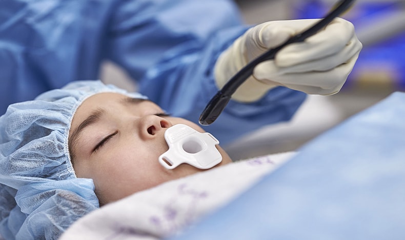
To achieve first-time-right imaging, a significant challenge that any of us who have worked with children will know is their fear of imaging procedures, which can increase motion and degrade image quality. We also need to think about how we can image gently – reducing exposure to radiation and contrast agents to reduce the risk of additional complications, which in pediatric patients can further impact on later development. Getting excellent image quality to render a first-time-right diagnosis can require patients to lie still for as long as an hour. As I have seen first-hand, kids during imaging procedures move – a lot – and so this is no easy task. This motion can affect image appropriateness and quality, and with 20% of rescans necessitated by motion artifacts, this is clearly a key issue. [2] This results in repeated scans and increased procedure times, costs, and patient radiation exposure.
Q: Children fidget and move, especially when they are anxious. How can we help them stay still to get the image quality right first time?
As I have seen first-hand, kids during imaging procedures move – a lot – and so this is no easy task. This motion can affect image appropriateness and quality, and with 20% of rescans are necessitated by motion artifacts [2]
This struggle to assist pediatric patients to lie still during imaging can be compounded by these kids’ anxiety around the procedures. In these cases, we may decide it is in the best interest of the patient to sedate them for imaging. But the use of sedation adds additional time, cost, and potential complications into the mix [3], as well as creating anxiety for parents. The most prevalent complication of sedation is cardiorespiratory depression with concomitant loss of protective reflexes, which may prove disastrous. [3, 4] And sedation may not always be successful. Sedated patients may still be agitated and unmanageable, resulting in failed imaging. [3] In fact, failures of adequate sedation are reported in 23% of cases. [5]
Supporting children with an interactive app and a child-friendly multimedia environment in the clinic reduced the need for anesthesia from 57% to just 5%. [6]
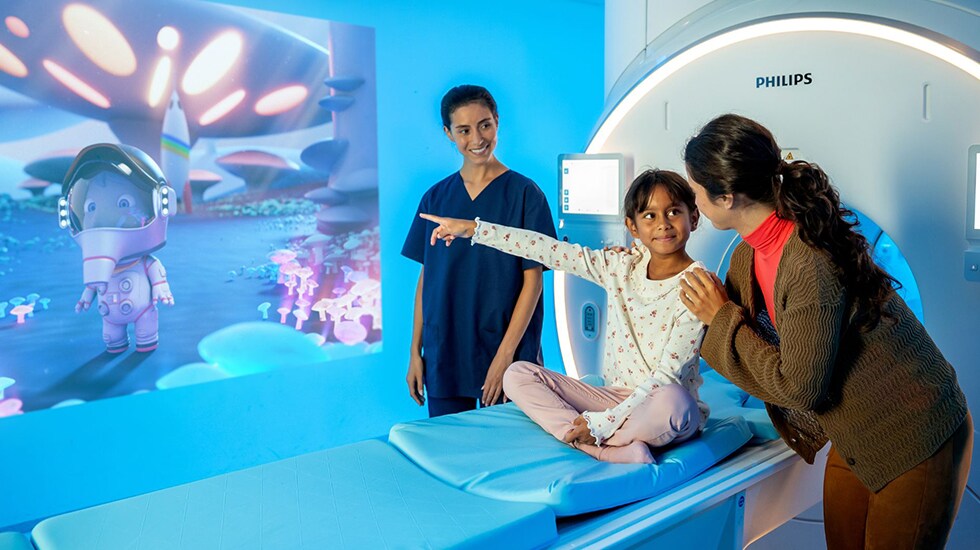
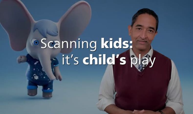
Researchers at Philips are taking a child-first approach to managing motion through ‘non-pharmacological sedation’ – using distraction, play therapy, and parental involvement as an alternative to anesthesia. [3] Through Philips’ Scan Buddy interactive app, kids can gain familiarity with the shape, sounds, and procedures of MRI ahead of their scan and from the comfort of their own home. From the Scan Buddy app through to the day of their appointment, Ollie the Elephant and his friends will coach them throughout their MRI experience. Children who feel more confident and in control are more at ease, improving the chances of a successful scanning procedure. [7] One study showed that supporting children with an interactive app and a child-friendly multimedia environment in the clinic reduced the need for anesthesia from 57% to just 5%. [6]
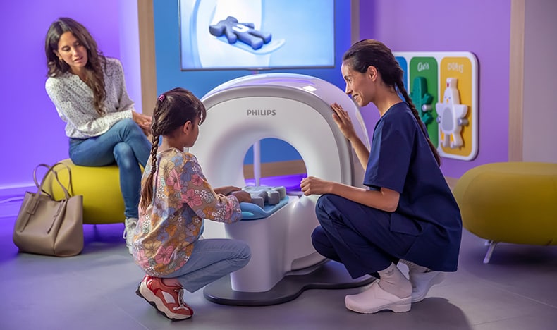
Along with Ollie, a comfortable and calming setting within the MRI room relaxes patients and reduces motion. Ambient Experience incorporates dynamic lighting, projection and sound to achieve this. Once patients are in the machine, In-bore Connect helps patients navigate breath holds and the overall duration of the exam. [8, 9, 10]
The number of interrupted scans was reduced by 70% with the use of Ambient Experience and In-bore Connect [10]
There is also a strong trend toward shorter exams. Indeed, the use of AI in software applications such as Philips’ SmartSpeed has been able to increase imaging speed by up to a factor of three with no compromise to image quality. [2] This is great news for pediatric and adult patients alike.

Q: Why is reducing exposure to radiation and contrast agents especially important for the safety of pediatric patients?
Primum non nocere. First do no harm. Every physician knows this quote well, and for pediatric imaging, this remains true. We must image gently. But radiation should be respected, not feared.
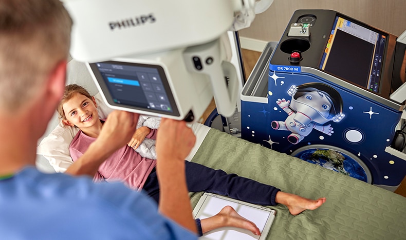
Primum non nocere. First do no harm. Every physician knows this quote well, and for pediatric imaging, this remains true. We must image gently. But radiation should be respected, not feared. When imaging, we must ensure radiation dose is ALARA – ‘As Low As Reasonably Possible’. Children are more at risk from ionizing radiation’s harmful effects versus older adults. Often, higher relative doses of radiation are required in pediatrics. Children typically will have many more decades of life following imaging with ionizing radiation compared with adults. And often they have repeated imaging. Thus the chance of side effects arising from the cumulative effects of radiation is increased. Studies show that the risks from radiation are around 10–11 times higher in pediatric patients than in adults. [7] Iodinated contrast and gadolinium are contrast agents that are used in CT and MRI which can prove toxic in some cases. But ALARA holds true here as well. Just like with radiation, contrast also should be respected and not feared: another reason why patient experience solutions like Ambient Experience that reduce the need for rescans are so clinically relevant. Let’s look at CT, while it is difficult to show that there is a direct link between CT scans and cancer, according to one landmark study from 2012 funded by both the United States’ National Cancer Institute and the United Kingdom’s Department of Health, having CT scans in childhood can triple the risk of leukemia and brain cancer later in life. [11] This reinforces the need for us to get ‘first-time-right’ diagnosis from pediatric CT exams. It’s no wonder that spectral CT imaging – which acquires multiple data sets simultaneously – is a popular option for pediatric cases. Compared to traditional CT, spectral CT reduces radiation and contrast dose requirements while allowing for improved image quality, providing an ‘image gently’ solution that decreases the need for follow-up evaluation. [12] We’re also seeing an encouraging trend with traditional CT systems such as Philips CT 5300, which has reduced radiation dose by as much as 80% thanks to its AI-powered Precise Image reconstruction technology.* [13, 14] Kids are not just small adults. They vary tremendously in size. As I have seen first-hand, we image infants as small as a few ounces to teens weighing over 200 lbs. It’s why we have developed specialized scanning protocols. Radiologists must balance the necessity of diagnostic imaging with the need to limit radiation exposure, and developing age- and weight-appropriate imaging techniques and adjusting radiation doses accordingly is essential. Acquiring high-quality pediatric images at a dose that is ALARA is possible only through the use of innovative technologies and workflow efficiencies. In diagnostic X-ray, the Philips Radiography 7000 M is a premium mobile digital radiography (DR) solution that supports innovative dose management and techniques for the acquisition of quality pediatric images. Kevin McDevitt, Diagnostic Radiographer, Western Health & Social Care Trust, expressed that: “The Philips DigitalDiagnost C90 has a camera on the X-ray tube which means we can check that our patient is still in the same position before we expose. We can adjust the collimation, if necessary, which helps to reduce the number of repeat exams and, in turn, the patient receives a lot less radiation exposure.”
Q: It’s one thing to provide better care for children who have access to quality care – but what about those children who don’t have access at all?
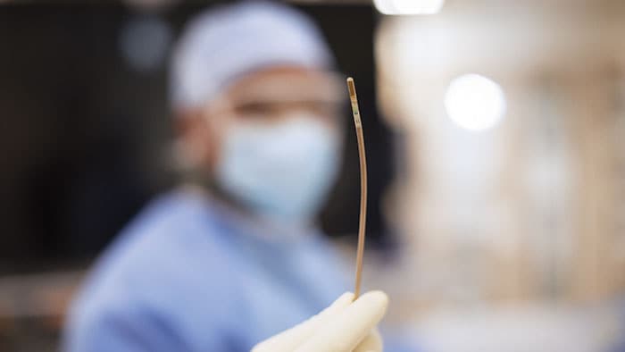
Lack of access to resources for pediatric care falls into two main areas of need: access to the technology itself, and access to clinical staff trained in the unique requirements of pediatric care. On the technology front, several trends are making a difference. AI, miniaturization, and cloud-based informatics are bringing care to more children every day. Take the example of our Lumify miniaturized hand-held ultrasound, which can be attached to a mobile phone or tablet. The Philips Foundation is working with local healthcare providers in the Philippines and in Uganda to screen children at risk for rheumatic heart disease (RHD). It’s helping providers identify at-risk patients and averting tragic complications that come with undiagnosed RHD. [15] Another example is Philips’ new Mini 3D TEE (transesophageal echocardiography) ultrasound transducer, used for the minimally invasive diagnosis and treatment of damaged heart valves and congenital heart defects. While a larger version has been around for a decade, only recently has miniaturization technology allowed pediatric patients as small as 5 kg to benefit from this valuable technology – with huge implications. [16] Using ultrasound in place of radiation-based imaging is a clear way to reduce imaging radiation risk for a patient. And having a probe that is 35% smaller than its predecessor allows even more children – in more places – to benefit from these life-saving procedures.
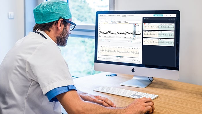
Even with access to imaging technology, scarcity in ultrasound scanning and interpretation expertise remains a problem. Thus, Philips researchers have been working with partners in Kenya and other low and middle-income countries, embedding artificial intelligence (AI) into our Lumify ultrasound. This project, partially supported by the Bill and Melinda Gates Foundation, is enhancing the skills of midwives and other providers to help them reduce maternal and infant morbidity and mortality. This type of technology aims to make high-quality prenatal care much more affordable in low-resource settings and prevent the pediatric complications of prematurity. [17] By using virtualization technologies such as Collaboration Live in our ultrasound systems and remote operation command centers (ROCC) for CT and MR systems, expert physicians and technologists can ‘beam in’, helping empower less experienced providers through even more complex exams. This is great news for pediatric patients in remote and underserved areas, who can now benefit from specialist expertise. [18]
Conclusion
Quite simply, by putting digital health tools together and connecting the dots across imaging, we can get even better at making sure that first-time-right diagnoses and treatments are available for every child. No matter who, no matter where.
Imaging children comes with unique challenges for my fellow radiologists and technologists. We need to obtain excellent image quality to enable first-time-right diagnosis. We strive to further reduce radiation and contrast to make scans even safer. We need to consider how we can deliver even more inclusive pediatric patient care, and for every child, increasing access to care. And we want to maximize patient safety and comfort, by putting the experience of each child first.
*In clinical practice, the use of Precise Image may reduce CT patient dose depending on the clinical task, patient size, and anatomical location. A consultation with a radiologist and a physicist should be made to determine the appropriate dose to obtain diagnostic image quality for the particular clinical task. Dose reduction assessments were performed using reference body protocols with 1.0 mm slices at the “Smoother” setting, and tested on the MITA CT IQ Phantom (CCT189, The Phantom Laboratory) assessing the 10 mm pin and compared to filtered-back projection. A range is seen across the four pins, using a channelized hoteling observer tool, that includes lower image noise by 85% and improved low-contrast detectability from 0% to 60% at 50% to 80% dose reduction. NPS curve shift is used to evaluate image appearance, as measured on a 20 cm water phantom in the center 50 mm x 50 mm region of interest, with an average shift of 6% or less.
Sources [1] A. Ferrari, et al. "The Sooner the Better? How Symptom Interval Correlates With Outcome in Children and Adolescents With Solid Tumors: Regression Tree Analysis of the Findings of a Prospective Study," Pediatr Blood Cancer, vol. 63, no. 3, pp. 479-85, 2016. [2] Philips, "Philips SmartSpeed. No compromise: Image quality and speed at your fingertips," Philips, 2022. [3] Y. Arlachov and R. H. Ganatra, "Sedation/anaesthesia in paediatric radiology," Br J Radiol, vol. 85, no. 1019, pp. e1018-31, 2012. [4] M. R. Moradkhani, et al. Drug anesthesia for children undergoing magnetic resonance imaging: A review," Biomed Pharmacother, vol. 88, pp. 1183-7, 2017. [5] S. Malviya, et al. "Adverse events and risk factors associated with the sedation of children by nonanaesthesiologists," Anaesth Analg, vol. 85, pp. 1207-13, 1997. [6] S. B. Runge, et al. "Children centered care: Minimizing the need for anesthesia with a multi-faceted concept for MRI in children aged 4-6," Eur J Radiol, vol. 107, 2018. [7] P. R. Regmi, et al. "Modern Paediatric Radiology: Meeting the Challenges in CT and MRI," JNMA J Nepal Med Assoc, vol. 60, no. 251, pp. 661-3, 2022. [8] Philips, "Patient comfort leads to first-time-right imaging," Philips, 2015. [Online]. Available: https://images.philips.com/is/content/PhilipsConsumer/Campaigns/HC20140401_DG/Documents/Fieldstrength_Ambient%20Experience_Herlev_Denmark.pdf. [Accessed 17 May 2024]. [9] M. van der Zwaag and H. Geraedts, Philips Research Laboratories Europe, 's-Hertogenbosch, The Netherlands, 2011. [10] Philips, "Ambient Experience for MR: Improving care and efficiency," Philip, 2017. [11] M. S. Pearce, et al. "Radiation exposure from CT scans in childhood and subsequent risk of leukaemia and brain tumours: a retrospective cohort study," Lancet, vol. 380, no. 9840, pp. 499-505, 2012. [12] S. Z. Adam, et al. "Spectral CT of the abdomen: Where are we now?," Insights Imaging, vol. 138, p. 12, 2021. [13] Philips, "CT 5300: Intelligence reimagined," Philips, [Online]. Available: https://www.philips.co.uk/healthcare/resources/landing/ct-5300. [Accessed 17 May 2024]. [14] Philips, "AI-enabled solutions," Philips, [Online]. Available: https://www.philips.com/a-w/about/artificial-intelligence/ai-enabled-solutions.html. [Accessed 23 May 2024]. [15] Philips Foundation, "Rheumatic heart disease screening program in Uganda," Philips Foundation, 2019. [Online]. Available: https://www.philips-foundation.com/a-w/knowledge-hub/2019-017.html. [Accessed 23 May 2024]. [16] Philips, "New Philips Mini TEE ultrasound transducer helps improve cardiac care for more patients," Philips, 31 January 2024. [Online]. Available: https://www.philips.com/a-w/about/news/archive/standard/news/press/2024/new-philips-mini-tee-ultrasound-transducer-helps-improve-cardiac-care-for-more-patients.html. [Accessed 23 May 2024]. [17] K. O'Reilly, "Philips program developing AI-powered ultrasound to expand access to maternal health receives major funding boost," Philips, 07 November 2023. [Online]. Available: https://www.philips.com/a-w/about/news/archive/standard/news/press/2023/20231107-philips-program-developing-ai-powered-ultrasound-to-expand-access-to-maternal-health-receives-major-funding-boost.html. [Accessed 23 May 2024]. [18] Philips, "Philips Collaboration Live," Philips, [Online]. Available: https://www.philips.co.uk/healthcare/resources/landing/collaboration-live. [Accessed 23 May 2024].
Media contacts

Contact details Contact details
You are about to visit a Philips global content page
Continue
Contact details Contact details
You are about to visit a Philips global content page
Continue