
Dec 13, 2021
Ultrasound rises to the challenges of 21st century imaging
From screening and diagnosis to treatment guidance and assessment, it is impossible to imagine healthcare without medical imaging. What would have been perceived as miraculous a century ago is taken for granted today: we can peer into the smallest vessels, watch the brain at work, observe fetal development and virtually travel through the digestive system. We can both pinpoint tiny tumors and image the entire body at once, and perform image-guided surgery without major incisions. Medical imaging professionals who appreciate the power of diagnostic images to guide care decisions and delivery are also aware of where imaging falls short. In image acquisition, there is often a wide gap between those expert operators who can reliably acquire pristine images, and those who struggle with consistency. In interpretation, radiologists can unlock all sorts of information from an imaging study, yet non-diagnostic studies still occur, and images aren't as accessible for non-radiologists. And around the world, many patients lack access to the imaging they need.
Ultrasound innovation bridges the gaps
We at Philips know we must bridge these gaps if diagnostic imaging is to reach its full potential. As we look to solve challenges in imaging standardization, diagnostic certainty, and access, we're finding answers in a somewhat unexpected place: ultrasound. Ultrasound has made progress in addressing its traditional weaknesses in operator dependency and image quality, and retains its advantages in affordability, portability, and lack of ionizing radiation.
Increased consistency regardless of operator
Ultrasound is historically one of the most operator-dependent medical devices. An experienced, expert technologist can coax remarkably clear images from a transducer, while a less experienced user might be unable to get diagnostic images from the best equipment. But today, advances in artificial intelligence (AI),* automation and live collaboration have countered this traditional challenge, making ultrasound now easier to use than ever. After an exam, AI can check the quality of the resulting images. This capability gives technologists immediate feedback, helping to promote confidence in their technique and helping to prevent callbacks.
In addition, AI enables automated measurements for a variety of applications, including abdominal, cardiac, breast and fetal imaging. For example, 3D imaging combines with new technology to provide user-independent measurements of abdominal aortic aneurysms. Removing potential variability from this important measurement is critical when managing aneurysm progression.
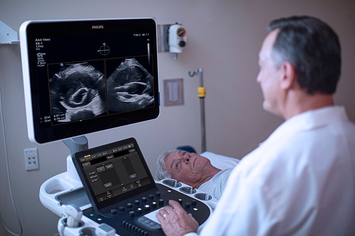
Abdominal aortic aneurysms (AAAs) cause more than 175,000 deaths globally every year, with an 80% mortality rate if ruptured.[1] Philips AAA Model is designed to segment and quantify 3D ultrasound data for use in surveillance of native and post-endovascular aneurysm repair (EVAR) AAAs.
AI also brings new efficiency to all aspects of post-processing, including choosing the best images for diagnosis, color-coding anatomy, and voice recognition technology. Automation partners with AI to increase consistency regardless of operator, while also reducing repetitive tasks. Technology that automatically optimizes gain and TGC assures that optimal images are achieved in 2D, 3D, or 4D. Philips SmartExam decreases exam time by 30-50%, keystrokes by as many as 300/exam, and results in a higher level of consistency among users. Auto Doppler takes time-consuming color box positioning and sample volume placement from ten steps to three steps and reduces the number of repetitive button-pushes by an average of 67.9%. Consistent imaging can help physicians assess severity of disease, plan treatment, and compare exams over time. All these advances also can increase staff satisfaction by freeing up staff time for their most challenging clinical assessments, and allowing technologists to focus their expertise and energy on the patient.
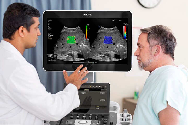
Greater diagnostic certainty
In the past, ultrasound's low-contrast resolution limited its application. While cost and convenience made it a common first modality, often imaging using other modalities was required for diagnostic certainty. The development of advanced imaging modes, including harmonic imaging, spatial compounding, and tissue aberration correction reduce artifacts and enhance contrast resolution, enabling ultrasound to deliver new insights across anatomies and applications. Today, ultrasound systems quantify liver fat, aid in detecting breast cancer that mammography may miss in women with dense breasts, deliver photo-realistic views, change the lighting source to backlight anatomical structures, and image blood flow in small vessels to check for cancer or inflammation or to assess perfusion, to name just a few examples.
Linked strongly with obesity, it is estimated that non-alcoholic fatty liver disease affects as many as one billion individuals worldwide. Philips Liver Fat Quantification (LFQ) tools provide an accurate and reproducible method of measuring liver fat.
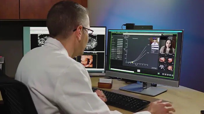
Advances also enable system operators to support diagnostic certainty by accessing expertise remotely. With Collaboration Live, ultrasound operators can connect with experts quickly and securely from the ultrasound system and receive guidance and support via Windows, Chrome, iOS or Android devices.
The future promises to further expand applications, driven by:
Greater accessibility
The World Health Organization (WHO) estimates that two-thirds of the world’s population lack access to basic medical imaging technology, causing preventable and sometimes fatal delays in diagnosis and treatment.[2] For many, access is limited simply because diagnostic imaging systems are often expensive and stationary, and transportation is inconvenient and limited.[3] The U.S. veteran in a rural location and the subsistence farmer in a developing country have the same challenge: imaging systems are only in large hospitals many miles from their homes. Ultrasound's greatest contribution in the future may lie in innovation that brings imaging to new locations. Even within a hospital, it can be difficult to bring very ill patients to the imaging department. During the ongoing Covid pandemic, physicians are using ultrasound imaging for bedside diagnosis, to avoid transporting patients to imaging departments. In fact, wireless transducers make it possible to leave the system itself outside the room, decreasing opportunities for cross-contamination. The use of ultrasound as a bedside imaging modality is likely to continue even when the pandemic is over. Ultrasound is also appearing more frequently outside the hospital, in doctor's offices. In the future, as ease of use increases, primary care physicians may reach for a multi-application transducer just as they reach for a blood pressure cuff, and perform a quick imaging study to determine if a patient should be referred to a specialist. New collaborative tools bridge the gap between operators and expertise so that ultrasound can be used in remote locations and for underserved populations. Collaboration Live uses tele-health technology to give system operators on-demand, secure access to expert physicians and technologists for real-time guidance and decision-support.
Staff limitations can make it challenging to deliver the full value of ultrasound to all who could benefit. Help close the gaps with Philips tele-ultrasound remote diagnosis capabilities.
In some locations, the cost of systems is a barrier to widespread use. Simpler systems, in which access to powerful off-system processing is available through the cloud, may enable healthcare systems and governments to place ultrasound systems in a greater number of locations. Already today, a transducer paired with a mobile device can convert a smartphone into an ultrasound system, combining two-way audio-visual calls with live ultrasound streaming. When we consider ultrasound's possibilities, it strengthens our resolve to push the limits of what this remarkable technology can do. Given the dedicated and talented clinical collaborators around the world who join us in this effort, we know that regardless of how far ultrasound has come, there are still new miracles before us. *According to the definition of AI from the EU High-Level Expert Group
1. Howard DP, Banerjee A, Fairhead JF, et al. Age-specific incidence, risk factors and outcome of acute abdominal aortic aneurysms in a defined population. British Journal of Surgery. 2015;102(8):907-915. doi:10.1002/bjs.9838. www.ncbi.nlm.nih.gov/pmc/articles/PMC4687424
2. Morris, M.A., Saboury, B. (2019). Access to Imaging Technology in Global Health. In: Radiology in Global Health, 15-33. https://link.springer.com/chapter/10.1007/978-3-319-98485-8_3.
3. Loria, Keith. Accessible Care: Challenges and Opportunities Related to Radiology Services in Rural Areas. Radiology Today. Vol. 20 No. 12 p .22. https://www.radiologytoday.net/archive/rt1219p22.shtml.
Share on social media
Topics
Author

Jocelyne Mekontso
Product Management Lead for General Imaging Ultrasound at Philips and the ad-interim leader for the Category Jocelyne brings more than 17 years of experience working in healthcare, in different locations such as the United States, Korea, Ghana and France, leading global product development, portfolio and P&L management, go-to-market strategy, indirect sales, program management and R&D. Along her career, Jocelyne performed program and product management roles in different segments such as mammography, general imaging ultrasound, diagnostic cardiology and global services.
Jocelyne has a Bachelor in Biology & Chemistry from Pierre and Marie Curie University, a MSc in Engineering Healthcare Information Technology from Polytech Grenoble. She is also a graduate of the GE’s accelerated leadership program (XLP). She speaks English and French fluently with elementary proficiency in Korean and Spanish.
Follow me on
Related news
-
![What will tomorrow’s smart hospital look like?]()
October 19, 2022
-
![Cardiac care without boundaries]()
July 22, 2021
-
![Why AI in healthcare needs human-centered design]()
April 19, 2021


