
Jun 02, 2021
Four breakthrough innovations in pediatric imaging: getting care right for the youngest patients
My close friend who works in the pediatric echo lab at the University of California, Los Angeles, recently celebrated the 17th birthday of her first heart transplant patient. As she told me about the celebration, I was moved beyond words – not just because I am also a parent myself, but because it’s the ultimate culmination of what we aspire to do to improve pediatric imaging. This journey toward a 17th birthday started with an ultrasound scan of this child’s heart more than a decade ago. These can be challenging scans because, as any parent knows, when children come to the doctor’s office they are scared, squirmy, maybe even crying. Parents are also anxious to get their kids back home, so any imaging procedure needs to be quick and done right the first time. And it needs to be done with specific tools that are tailored to the unique anatomies of younger patients. Kids aren’t just small adults, after all. Innovations in ultrasound and echocardiography since that initial scan have no doubt led to more stories such as this one. And across pediatric care, breakthroughs in other methods of imagining are helping provide the best possible care for our youngest patients. Improving pediatric imaging is critical because it’s increasingly popular today. Healthcare organizations’ investment in pediatric imaging exceeds $6B worldwide and is projected to grow 7% by 2027.[1] Higher incidences of premature births, pediatric diseases, and injuries (all requiring specified imaging devices), as well as more demand for preventive care, are all pushing the need for continued innovation in pediatric imaging. As a sonographer with an abiding interest in our youngest patients, I truly believe that the pediatric imaging industry is up for the challenge. In fact, I see the industry already innovating to meet this demand by developing new and unique pediatric imaging systems and tools that can quickly, safely, and precisely visualize and diagnose complex conditions in pediatric patients based on their unique needs. Here are four innovations that I’m most excited about.
Detecting heart disease in utero
One of the most sophisticated advances in pediatric imaging is the ability to detect fetal anomalies in utero – especially heart defects related to congenital heart disease. About 1 in 100 babies is born with congenital heart disease, and a third of these children will undergo surgical or catheter-based intervention in the first year of life.[i] When caught early, kids have a higher chance of celebrating more birthdays than if these defects are not found until later in life.
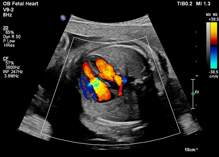
Ultrasound image demonstrating a fetal heart at 28 weeks We are continuing to innovate in this area and have even developed solutions that incorporate MRI to address congenital abnormalities in the womb. This fetal cardiac imaging innovation is helping to image complicated pathologies before a baby is even born. This can allow treatment planning to begin while the baby is in the womb, giving them the best chance at the best possible outcome. These innovations are so exciting because if an anomaly is detected early, some surgeries today can even be done in utero to fix heart defects, which improves outcomes because kids recover better while still immersed in amniotic fluid.[i],[ii],[iii] Catching cardiac abnormalities early also helps prevent complications at birth because moms can then plan to give birth at hospitals that are equipped to handle any expected complications, which gives their baby the best chance at survival.
If a prenatal ultrasound finds something that warrants further investigation, or if the fetus is at a higher risk for a heart defect, then fetal heart echocardiography is warranted to screen for these defects. Fetal echo, which uses transducers and applications designed specifically to detect anomalies in the fetal heart, is used for the diagnosis of the most complicated congenital anomalies. During the fetal echo, clinicians can evaluate the structure and function of the baby’s heart, examining an almond-sized object beating about 150 times per minute, for small holes and connections. As the technology has matured, we’re seeing innovations in fetal echo create higher accuracy scans, while also optimizing the workflow so that the transducers are easier for clinicians to use.
Reducing pediatric patients’ fear and anxiety
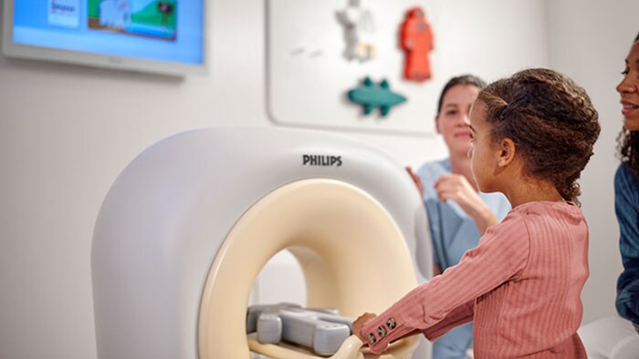
For pediatric patients and their parents, imaging examinations can be one of the most frightening experiences in their care journey. This poses a challenge for medical staff because they need to take great care to help patients stay calm and still during the scan. Yet too often, this isn’t possible. This leads to repeated scans, which happens in an estimated 20-30% of scans,[i] or to the need for sedation, which may pose long-term health risks, such as impaired brain development.[ii] Improving the pediatric MRI experience is an area that is filled with innovation. Because MRI doesn’t involve ionizing radiation, it’s becoming a preferred method of scanning for diagnosing a wide range of conditions in children due to injury, illness, or congenital abnormalities; these range from evaluating injury after trauma to monitoring response to cancer treatment or surgery. Today, more than 80% of MRI sites perform pediatric MRI procedures, and one out of 10 MRI examinations is a pediatric exam.[iii] To support this need, innovations in MRI techniques specifically for pediatrics is creating a much more kid-friendly experience, which is leading to better outcomes. To help reduce a child’s fear about MRI examinations, we’re building on the success of our Ambient Experience solutions for MR to create an end-to-end experience that helps children prepare for their exam in advance. For this purpose, we developed an award-winning app and coloring book that helps educate children about the experience in advance. The app uses videos and gamification to give children an idea of what to expect. Once at their appointment, they can engage with the KittenScanner, which is a toy scanner that playfully educates them about the scanning process. Once inside an MRI system, the Philips Ambient Experience provides an immersive video experience that distracts and entertains pediatric patients while lying in the bore.
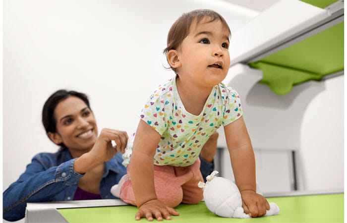
Before undergoing an MR exam, children can play with an educational app (at home) and a toy scanner (in the hospital) to get an idea of what to expect Pediatric patients often experience high levels of stress in uptake rooms, such as when oncology patients visit the hospital for a PET/CT scan. We’ve also worked closely with Phoenix Children’s Hospital to develop an Ambient Experience environment in PET/CT uptake rooms that calms patients to help relieve their stress, which can lead to better imaging results. In an exciting potential development for pediatric imaging, Philips recently partnered with the Walt Disney Company EMEA on a research collaboration to test custom Disney themes within Philips Ambient Experience in select hospitals across Europe. If the project moves into a commercial phase, MR patients would be able to select their favorite Disney story and character to provide them with a sense of control and comfort.
Fighting pediatric obesity
Sadly, childhood obesity has become a global epidemic. This has led to children as young as 8 years old presenting in the clinic with fatty livers, altered liver function, and even Type II diabetes. What was once known as adult-onset diabetes is now turning up more and more in childhood. This is a troubling trend, but if we can find signs of liver disease early enough, start treatment and monitor progression, we have a better chance at reversing the damage by stopping the cause.
Special ultrasound technology, designed specifically for pediatric care, has been a breakthrough innovation to monitor pediatric liver health. We’ve created an ultrasound solution designed for pediatrics, for example, that is tailored to provide quick and confident, yet gentle imaging specific to the needs of children, elevating their care as never before. With specialty transducers and elastography optimized for children, it delivers a 30% improvement in penetration when compared to the previous generation of pediatric transducers.[i] It also has functionality that provides real-time large field of view assessment of tissue stiffness, which is ideal for the pediatric patient who may be challenging to image; this innovation may prevent the need for liver biopsy. At Phoenix Children’s Hospital, this solution for whole liver imaging is helping kids improve their liver health.[ii] For example, an 11-year-old overweight male with elevated liver enzymes and concerns for non-alcoholic steatohepatitis (NASH) was referred for ultrasound of the liver with Doppler and Shear Wave Elastography. The ultrasound revealed signs of hepatic steatosis, an accumulation of fat in the liver that could lead to NASH. The patient could then be referred to a registered dietician for weight loss without the need for further imaging or biopsy.
Better image quality and outcomes for the most common imaging experience
Radiography is still the most common imaging experience for pediatric patients. Having an X-ray performed for a possible broken bone is almost a rite of passage for children. But because it is so common and widely available, it’s important to make sure that radiographic examinations are as safe as possible. Radiographic examinations expose kids to radiation, so they need to be done right the first time to keep radiation dose as low as possible. I’m happy to see pediatric-specific innovations helping deliver a calm, safe and accurate X-ray experience. We’ve designed our portfolio of digital radiography and fluoroscopy systems for pediatric patients from the ground up. Grid controlled fluoroscopy for example is an imaging technique unique to Philips that allows for dose reduction by real-time dose adjustment via the capacity to adjust the intensity of each pulse in real-time. Additional features such as automatic filters and collimation on last image hold, were designed to benefit both the young patient and the staff.
Radiography, or X-ray, is still the most common modality in pediatric imaging and can be administered in a way that delivers a calm and safe experience even for the youngest patients Another example of a designing for pediatrics is a tube head that was designed with pediatric patients in mind. By incorporating a large touchscreen monitor, technologists can work right next to the patient, helping calm their nerves. And before taking an exposure from the control room, the technologist can verify that the patient hasn’t moved thus avoiding unnecessary retakes. Other innovative tools are helping drive workflow efficiency in radiography, allowing technologists to see more patients per day and shorten wait times by decreasing the time to diagnosis. For example, because a pediatric patient ranges from newborn to 18-year-old, defining the patient properly for protocol adherence can be a time-consuming task. The interface on our digital radiography systems allows technologists to quickly select from multiple different patient profiles. There are three profiles for pediatric patient that the system can determine automatically according to date of birth. Each profile optimizes for low dose and high image quality for that specific patient.
Pediatric imaging done right: giving kids the best chance at the best outcomes
Seamless care that is designed for the pediatric patient is what makes milestones like the 17th birthday of my friend’s heart transplant patient possible. It’s what drives me to push for intentional innovation every day so that more pediatric scans can be done right the first time to deliver the most diagnostic value with the least risk to our youngest patients. Realizing this vision requires a deep commitment to research and development in pediatric imaging, so that every pediatric patient has an imaging experience that is safe, comfortable and precise. Each pediatric imaging procedure has its own risks, benefits, challenges and potential, which is why their optimization for pediatric patients must be constant. Children have a lifetime in which they will either derive benefit from or be harmed by imaging choices made on their behalf. We owe it to them to create technologies and experiences that produce the best possible outcomes.
[1] https://www.grandviewresearch.com/industry-analysis/pediatric-imaging-market#:~:text=b.-,The%20global%20pediatric%20imaging%20market%20size%20was%20valued%20at%20USD,USD%207.3%20billion%20in%202020.&text=The%20global%20pediatric%20imaging%20market%20is%20expected%20to%20grow%20at,USD%2012.1%20billion%20by%202027. [i] https://www.heart.org/en/news/2018/12/17/halfway-through-pregnancy-their-baby-needed-in-utero-heart-surgery-should-they-risk-it [ii] https://give.brighamandwomens.org/fetal-cardiac-surgery/ [iii] https://www.nationwidechildrens.org/specialties/fetal-cardiac-program/fetal-cardiac-intervention [i] https://journals.sagepub.com/doi/full/10.1177/0218492317694248 [i] 20 – 30% of scans need to be repeated due to motion (Field strength MRI Magazine, June 2016) [ii] https://www.ncbi.nlm.nih.gov/pmc/articles/PMC6078876/ [iii] IMV 2019 MR Market Outlook Report [i] *Internal measured comparison on calibrated tissue phantom between mC12-3 and C8-5 transducers on EPIQ Elite ultrasound system. [ii] “Diagnosis and management of pediatric steatosis using ultrasound,” Philips Clinical Case Study, 2021
Share on social media
Topics
Author

Dr. Julia Dmitrieva, RDMS, RDCS, RVT, FSVU
KOL Engagement Leader for Precision Diagnosis at Philips Julia is passionate about providing the highest quality of care and improving patient experience. She is dedicated to creating and propagating solutions that improve clinical and operational outcomes and is an expert in ultrasound imaging.
Follow me on
Related news
-
![AI is changing how care is delivered today]()
June 11, 2026
-
June 11, 2026
-
![The race against time: how MRI innovation is transforming Alzheimer's care]()
September 19, 2025
-
![Equitable access to care for mothers and children]()
December 11, 2023
-
![Making the case for systemic change in healthcare]()
October 25, 2023
-
![Equitable access to care for mothers and children]()
September 08, 2022
-
![Healthcare has long been a place you go to. What if it came to you instead?]()
September 05, 2022
-
![Three ways the private sector can help build health system resilience]()
September 15, 2021


