Independent study shows dual-layer spectral CT performs among the best in head-to-head comparison with photon-counting CT
Mar 24, 2026 | 3 minute read
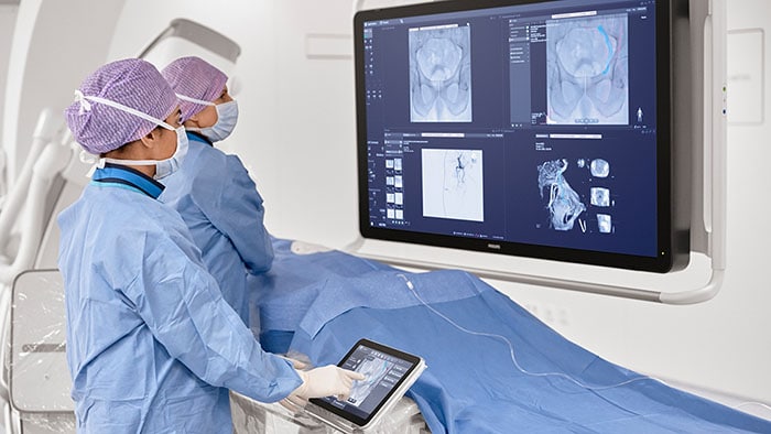
Across emergency, oncology, vascular and cardiac imaging, clinicians often face the challenge of clearly visualizing subtle contrast differences, while keeping CT scanning protocols efficient and consistent. Spectral CT imaging helps address this need by delivering clinicians additional information not only about anatomy but also spectral characteristics of tissues, which is designed to support visualization and assessment of lesions.
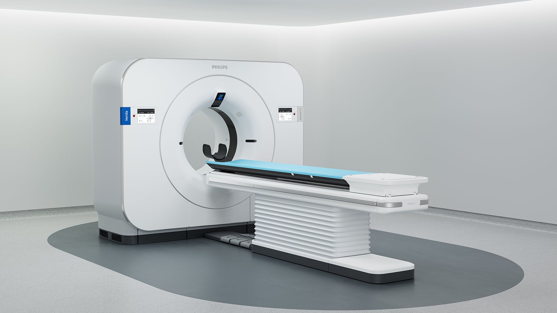
Detector-based spectral CT, also known as dual-layer CT (DLCT), can capture low and high energy X-ray information simultaneously using a two-layer detector. The scanner collects spectral data during every scan, enabling particular reconstructions called virtual monoenergetic images (VMIs) from 40-200 kiloelectron volts (keV), an energy range particularly important for contrast-enhanced imaging, along with iodine maps all without the need for a special scan mode or advance planning. This means the scanner can reconstruct images at different (in this case lower) X-ray energy levels as required and generate data that shows where the iodine contrast dye is in the body. This helps doctors highlight blood flow and provides detailed imaging information. In practical terms, this approach helps imaging teams maintain consistent CT protocols while still having access to spectral information when diagnostic insight is needed.
Independent study evaluates spectral CT performance
A recent independent phantom study [1] published in Diagnostic and Interventional Imaging compared the spectral performance of several advanced CT technologies, including two dual-energy CT (DECT) systems, one DLCT (the Philips dual-layer Spectral CT system) and one photon-counting CT (PCCT) system [1]. The study evaluated performance across low energy VMIs (40-70 keV) and iodine maps and assessed image-quality, modeled lesion visualization and iodine quantification across scanners with a blinded radiologist review.
Strong performance at clinically relevant low-energy levels
In the study, the DLCT system demonstrated some of the strongest performance [1] in the low-energy levels that matter most for contrast-enhanced imaging. Researchers found that lesion visualization at the clinically relevant low-energy range (40–50 keV) was highest for the DLCT system and the photon-counting CT system [1]. At these low energy levels, iodine appears brighter relative to surrounding tissues. This improved contrast can help clinicians better visualize subtle lesions.
Balanced image quality and reliable spectral measurements
In addition to lesion visualization, the researchers evaluated several image-quality characteristics that influence how clearly anatomical structures appear on CT images. These included image noise, noise texture and spatial resolution, assessed using standardized task-based image quality methods.
Results showed that the dual-layer spectral CT system delivered strong low-energy imaging performance while maintaining balanced image quality characteristics. In the blinded evaluation, radiologists also ranked images produced at 40–50 keV with dual-layer spectral CT among the most favorable overall.
The study also assessed the accuracy of iodine concentration measurements on iodine maps. Accurate measurement of iodine concentration in lesions may help support assessment of subtle lesions in clinical imaging. Across the systems evaluated in the study, iodine quantification accuracy for DLCT was among the strongest reported [1], which may support lesion visualization and characterization.
Independent validation of spectral CT capabilities
Although phantom studies cannot fully replicate clinical imaging conditions, they provide a standardized way to compare imaging technologies and quantitatively evaluate image quality.
The findings add to 1000+ peer-reviewed research papers supporting the clinical value of detector-based spectral CT imaging, particularly for contrast-enhanced examinations where improved iodine visibility can support lesion visualization and characterization.
Philips continues to advance spectral CT innovation, combining detector-based spectral imaging with AI reconstruction across the entire imaging chain, designed to support precision diagnosis and efficient clinical workflows.
Sources [1] https://www.sciencedirect.com/science/article/pii/S2211568425002128
General Disclaimer: Phantom studies provide controlled comparisons of imaging performance and may not fully represent clinical imaging conditions.
Media contacts

Contact details Contact details
You are about to visit a Philips global content page
Continue