Philips introduces first-of-its-kind multi-contrast 4D MR imaging for radiotherapy simulation
May 19, 2026 | 4 minute read
New 4D MR-radiotherapy (RT) solution combines respiratory motion-resolved 4D MRI with multiple image contrasts in a single free-breathing workflow, designed to help clinicians better visualize moving abdominal anatomies and improve patient comfort.
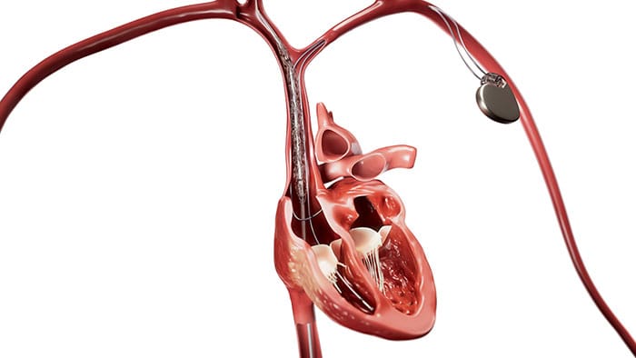
Amsterdam, the Netherlands – Royal Philips (NYSE: PHG, AEX: PHIA), a global leader in health technology, today introduced Philips 4D MR-RT, a new MR solution designed to improve visualization of abdominal anatomies affected by respiratory motion during radiotherapy simulation. The solution enables patients to breathe normally during imaging while supporting respiratory motion-resolved MR visualization of moving anatomies such as the liver and pancreas. Philips 4D MR-RT combines advanced 4D MR imaging with multiple image contrasts to help clinicians visualize moving anatomies and soft tissues more clearly.
A breakthrough in multi contrast 4D imaging for MR simulation
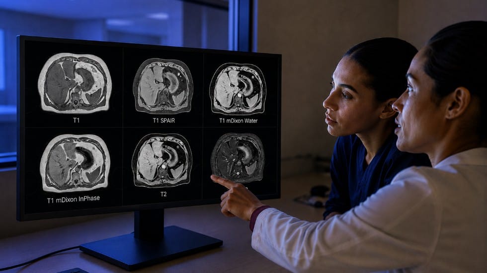
Different MR image contrasts can help clinicians visualize anatomical structures, soft tissues and lesions in different ways during radiotherapy simulation. Philips 4D MR-RT introduces a new multi-contrast 4D imaging with both T1 and T2 imaging – two complementary MR image contrasts that help clinicians visualize different tissue types and anatomical structures more clearly – with optional fat suppression during MR simulation of abdominal anatomies.
“Clinicians continue to look for imaging approaches that can help them better visualize abdominal anatomies affected by respiratory motion during radiotherapy simulation where movement during breathing can make treatment planning more challenging,” said Ioannis Panagiotelis, PhD, Business Leader, Magnetic Resonance at Philips. “With 4D MR-RT, Philips is advancing MR simulation, bringing greater confidence, consistency and accuracy to treatment planning, while supporting a more comfortable and reproducible patient experience. This marks another pivotal step toward Philips’ vision of self-driving MR."
Supporting clearer visualization during free-breathing abdominal MR simulation
Respiratory motion remains a challenge during abdominal MR simulation because patient breathing can blur abdominal structures and organs at risk, making it more difficult for clinicians to clearly visualize moving anatomies during radiotherapy simulation. While techniques such as breath-hold or compression may reduce such respiratory motion, they can adversely affect patient comfort.
Enabled by SmartSpeed, Philips 4D MR-RT supports respiratory motion management during abdominal MR simulation by distinguishing between inhale and exhale phases during normal breathing. This enables reconstruction of up to 10 respiratory phases to support clearer visualization of moving anatomies during free-breathing imaging.
Additional outputs, including separate respiratory phase images, mid-ventilation images and mid-position images, can also be generated to support contouring workflows for a range of Linac treatment strategies.
Philips 4D MR-RT is cleared for clinical use in the U.S. and Europe after receiving a CE Mark and 510(k) clearance.
The introduction of 4D MR-RT expands the Philips MR-RT portfolio with multi-contrast imaging capabilities for MR simulation. Learn more about Philips 4D MR-RT here.

Media contacts

Contact details Contact details
You are about to visit a Philips global content page
Continue