Seeing before scanning: Philips’ predictive preview on the path to autonomous MRI
Powered by NVIDIA accelerated computing technology and domain-specific foundation models, Philips is working to enable predictive MRI image generation to support smarter scan planning and more autonomous imaging workflows
Mar 17, 2026 | 6 minute read
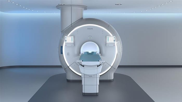
In today’s MRI suite, some of the most critical moments happen before the scanning even starts. A technologist reviews the referral, chooses a protocol, positions coils, aligns slices, and tries to anticipate the variables no checklist can fully capture – patient motion, unusual anatomy, metal artifacts, breath-hold challenges. Get the plan right, and the scan is smooth and the diagnostic information captured helps to provide a clear diagnosis. Get it wrong, and the cost is felt in rescans, delays, and uncertainty.
Now Philips is working to make that critical preview moment smarter using NVIDIA accelerated computing and open-source AI models. By generating an AI-driven image preview before data acquisition begins, the system can help validate scan plans and guide setup decisions. It’s part of a broader effort to move MRI toward a more autonomous future – where high-quality exams can be delivered consistently across operators, sites, and patient populations.
Teaching AI the language of MRI, from anatomy to artefacts
AI in medical imaging is often associated with algorithms trained for one task – detect a lesion, segment a structure, flag a scan for review. What Philips aims to build with NVIDIA has a different ambition. Their collaboration centers on a domain-specific MR Foundation Model trained on large-scale, diverse MRI datasets spanning anatomies, field strengths, protocols, and contrast settings. The aim of this MR foundation model is to interoperate with NVIDIA’s NV‑Generate‑MR, NV‑Segment, and NV‑Reason models, so that image generation, segmentation, and interpretation can be orchestrated as part of a single intelligent workflow.
Foundation models learn broad patterns from vast, heterogeneous data, producing generalized representations that can later be fine-tuned for specific applications. In MRI, that means teaching a model to understand the ‘language’ of MR across many conditions – how anatomy changes from head to toe, how sequences behave at different field strengths, how contrast and protocol choices shape appearance, and how artifacts arise.
NVIDIA accelerated computing and AI infrastructure provides the foundation to train and scale this kind of model. Building on Philips’ ongoing collaboration with NVIDIA – first announced in May 2025 (Philips collaborates with NVIDIA to improve patient care in MR with latest AI advances), Philips brings MR domain expertise and the intent to integrate AI capabilities directly into scanner workflows – not as a separate step, but as part of how an exam is planned, validated and executed.
The result will be an MR foundation model designed for scanner-integrated applications – and one of the most intriguing will be the ability to generate a predictive image preview before acquisition.
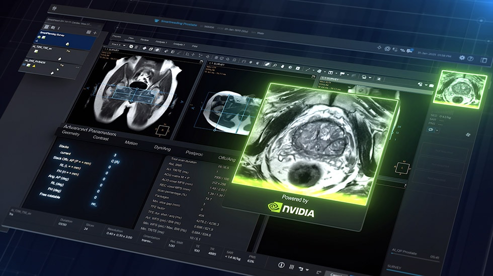
The breakthrough: seeing the scan before you scan
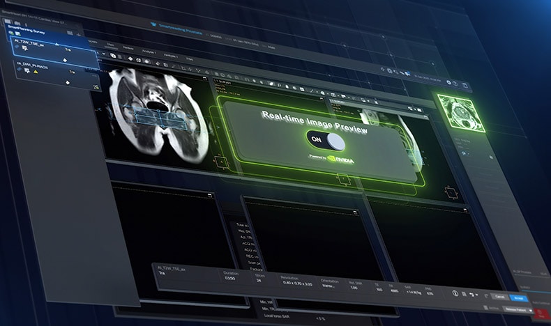
Imagine selecting an MRI protocol and, instead of waiting for the scanner’s first ‘localizer’ – the quick ‘setup shot’ technologists use to check they’re lined up correctly – the system gives you an intelligent first look straight away. This predictive preview is generated from patient context, the chosen protocol settings and the model’s learned understanding of anatomy.
The preview isn’t meant to replace the images the scanner actually acquires. Think of it as an informed prediction: a planning aid that will help the operator validate choices, fine-tune positioning and spot potential issues before the scanner commits time and resources.
For technologists and radiology teams, this seemingly small shift can make a real difference. MRI is incredibly powerful, but it can also be complex – and complexity is where variability creeps in. A predictive preview aims to reduce uncertainty at the moment it can cost the most: right before the scan begins.
Sathish Kumar Balakrishnan leads Research and Development for MRI at Philips: “By combining our MR foundation model with NVIDIA’s NV‑Segment for automated contouring, NV‑Generate for predictive previews, and NV‑Reason for context-aware decision support, we see an opportunity to make scan setup feel more guided and consistent for technologists,” says Sathish Kumar Balakrishnan. “For technologists and radiology teams, this seemingly small shift can make a real difference. MRI is incredibly powerful, but it can also be complex – and complexity is where variability creeps in. A predictive preview aims to reduce uncertainty at the moment it can cost the most: right before the scan begins.”
Philips is exploring how this image preview before scanning could help teams in a few practical ways:
Why autonomy in MRI matters now
The promise of autonomous MRI can sound futuristic until you consider the pressures facing healthcare systems today. Demand for imaging is rising, while many regions face staff shortages, burnout, and limited access to expert operators. Patients can wait weeks for appointments, and delays in diagnosis can mean delays in treatment.
An autonomous MRI system can simplify complex decisions, reduce variability, and support consistent, high-quality exams at higher volumes. If more sites can run reliable MR exams, more patients can be diagnosed earlier, including in primary care settings where early recognition of chronic and complex conditions can change the trajectory of disease.
That’s the underlying aim: helping more patients get diagnosed better and earlier, while easing the burden on staff and healthcare systems. Earlier answers can lead to earlier, more appropriate treatment – improving outcomes, shortening waiting lists, and reducing costs.
In Philips’ vision, the ‘gold standard’ for diagnostic imaging becomes autonomous MR – head-to-toe precision diagnostics available for every patient, in any location, from a megacity to a remote island.
Balakrishnan again: “The technology pieces are converging to make our ambition of autonomous MRI more than a slogan. We’ve already been laying groundwork through automation and integrated MR workflows, including a Dual AI engine designed to make scans faster while enhancing quality. And with our leadership in helium-free scanners, systems can be installed more sustainably and in places where traditional infrastructure made MR difficult – supporting resilience after disruptions and expanding access.”
Predictive image preview is a logical next step: improving the front end of the exam, where planning choices determine downstream success.
Dr. Ioannis Panagiotelis leads Philips’ MRI business: “Our continued collaboration with NVIDIA reflects our shared ambition to advance AI innovation in MRI using powerful models such as NV‑Segment, NV‑Generate, and NV‑Reason,” says Dr. Ioannis Panagiotelis. “By exploring foundation models trained specifically for MR, we aim to unlock new capabilities that could support workflow efficiency, consistency and operator confidence.”
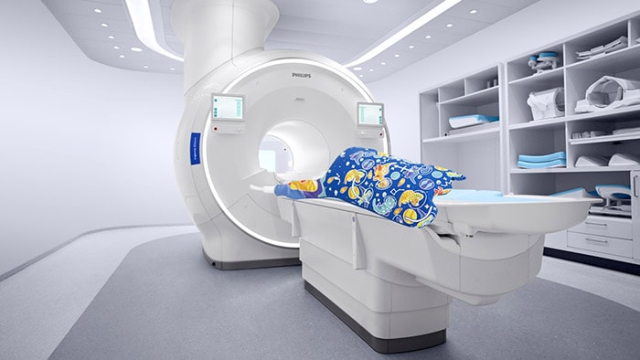
A patient’s experience in the near-future MRI
This is the kind of experience Philips is working towards as MRI becomes more intelligent and more automated. For patients, autonomy should feel less like robotics and more like reassurance.
Picture a future MRI visit that feels smoother, simpler and more predictable for everyone involved: a patient arrives for a scheduled exam and checks in at an automated kiosk. The system securely retrieves relevant information from the hospital record to prepare for the scan. Using integrated sensors and AI, the autonomous MRI automatically positions the patient and selects the appropriate scan, tailoring it to the individual and providing simple on-screen guidance.
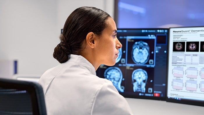
Before the scan begins, the system generates an intelligent preview – a quick, predictive look that helps validate the protocol and positioning. The scan then runs with AI continuously monitoring image quality and adjusting as needed to preserve diagnostic clarity. Once complete, the system helps the patient exit safely and flags any quality considerations for the radiologist, who is supported by AI-assisted insights during interpretation.
It’s a future where the most sophisticated imaging modality becomes more accessible, more consistent, and less dependent on scarce expertise – without compromising the clinical rigor that makes MRI valuable.
Self-driving MRI won’t arrive as a single leap. It will come as a series of practical steps that remove friction from the workflow, reduce variability, and improve outcomes. A pre-scan image preview – seeing the scan before you scan – is a major step on that journey.
Media contacts

Contact details Contact details
You are about to visit a Philips global content page
Continue