How KMC Manipal serves more patients with AI-enabled CT workflows
By Philips editorial team ∙ Jun 12, 2025 ∙ 3 min read
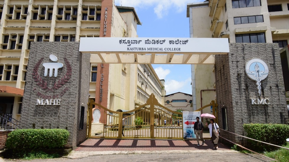
As part of a long-standing innovation partnership with Philips, the radiology department at Kasturba Medical College (KMC) Manipal in India implemented CT Smart Workflow in their CT exams and played a critical role in its clinical validation. This increased workflow efficiencies, allowing KMC Manipal to scan more patients daily, with better diagnostic confidence and improved patient experience.
Case study at a glance
Partner
Kasturba Medical College (KMC) Manipal, India
Challenge
With a growing demand for CT studies, the radiology department at KMC Manipal aimed to improve their workflows to serve patients and referring physicians more efficiently and effectively.
Solution
KMC Manipal implemented CT Smart Workflow, which includes AI-enabled tools to optimize various steps in the CT workflow, from patient positioning to image reconstruction and batching.
Results
By implementing CT Smart Workflow, KMC Manipal was able to save time on manual tasks, increase patient throughput, boost diagnostic confidence, and improve the overall patient experience.
The challenge
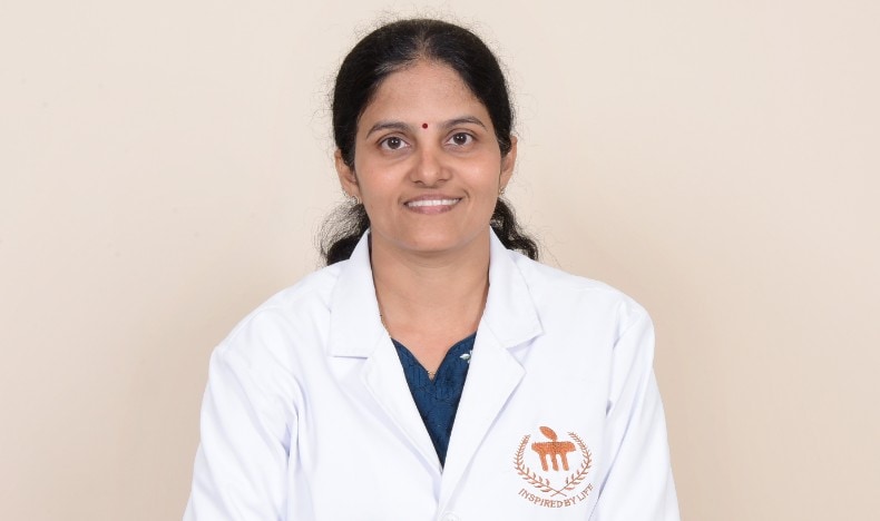
Like many radiology departments worldwide, KMC Manipal faces increasing patient demand, putting pressure on radiologists and imaging staff to reduce diagnostic turnaround times while maintaining high-quality patient care. This pressure is particularly felt in CT imaging due to its critical role in quickly diagnosing a wide range of medical conditions, including emergencies requiring immediate intervention. Any workflow inefficiency can lead to a loss of valuable time, requiring staff to work longer hours or risking delays in diagnosis and patient care. “Prior to implementing CT Smart Workflow, several steps in our CT workflow took more time than we wanted,” says Dr. Rajagopal K.V., Professor and Head of the Radio Diagnosis Department at KMC Manipal. Patient positioning was one such area. Dr. Prakashini K., Professor and Radiologist at KMC Manipal, explains: “We spent significant time training our medical imaging students to operate our CT machines and apply various clinical protocols. Each year we welcome new students, requiring ongoing training. While this is important, it can also be time-consuming, especially when we have heavy workloads. Patient positioning accuracy was not always optimal because it involved a lot of manual handling." The speed of image processing also left much to be desired, Dr. Rajagopal adds. “For example, batching brain scans required several manual steps, while time is of the essence, especially when we are dealing with trauma patients. In addition, images could be noisy, making it harder to notice subtle abnormalities. It was also a priority for us to minimize radiation dose for patients without compromising image quality, as many of our patients – such as those receiving cancer treatment – must undergo repeated scans.”
The approach
Building on a long-standing innovation partnership with Philips, KMC Manipal was one of the first hospitals to adopt CT Smart Workflow. Radiologists, PhD, MSc and BSc students at KMC Manipal also played a critical role in its clinical evaluation, ensuring its effective implementation while supporting cutting-edge academic research on its real-world benefits. Using the power of AI, CT Smart Workflow provides the radiology department at KMC Manipal a comprehensive suite of applications to optimize CT workflows at every step. Starting at the point of image acquisition, Precise Position automates patient positioning through an AI-enabled camera designed to increase positioning accuracy and user-to-user consistency. Radiologists at KMC Manipal also benefit from Precise Image, an AI-based reconstruction technique that uses the power of a deep-learning neural network for improved clinical confidence. Other applications that KMC Manipal’s radiologists use in their daily work include Precise Brain, which supports automatic batching of brain scans for timely analysis, and Precise Cardiac, which corrects for motion in cardiac images to improve image quality at high heart rates. “We already had a strong collaboration with Philips in CT, so implementing CT Smart Workflow was a natural next step for us,” reflects Dr. Prakashini. “The implementation went smoothly and was completed in just a few weeks. Having a dedicated innovation manager and clinical scientist from Philips on site has been key to our partnership. The user-friendly interface of the applications allowed our imaging staff and radiologists to get accustomed to them quickly. We have been happy with the expert support from Philips throughout the process. We have also teamed up on various research studies to demonstrate the benefits of CT Smart Workflow in clinical practice.”
Having a dedicated innovation manager and clinical scientist from Philips on site has been key to our partnership.
The results
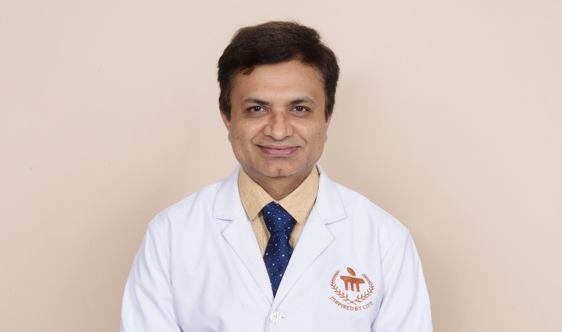
Three years into the use of CT Smart Workflow, the results have been significant. “With CT Smart Workflow, we have been able to achieve time savings throughout our entire workflow,” says Dr. Prakashini. “We used to do 70-80 exams a day, sometimes working until late at night to manage the case load. Now we scan 90-100 patients a day, with less overtime from staff. Referring clinicians are pleased that we are able to provide them with our images and reports more quickly, which helps to improve patient care.” Time savings start with patient positioning, Dr. Prakashini explains. “Previously, staff had to manually identify bodily landmarks and then correctly center the patient in the gantry by turning on the laser light and adjusting the region of the scan to the iso-center. But now, with Precise Position, that happens automatically with the press of a button. This can easily save us 1-2 minutes per case and helps consistently position even our most challenging patients, ensuring the region of interest is at the iso-center for optimal image quality at the optimal radiation dose.” She highlights additional time savings in areas such as automatic batching of brain scans using Precise Brain, which has saved up to 5 minutes per scan. Dr. Rajagopal adds how his department has benefited from improved image quality in many types of exams – including brain, chest, and abdomen – while simultaneously lowering radiation dose. “With Precise Image, we have seen significant reductions in image noise even at low dose, which helps us better detect subtle lesions or signs of metastasis in cancer patients,” he says. “Better image quality adds to our diagnostic confidence, which helps us deliver better care to patients. And by keeping dose levels as low as reasonably achievable, we avoid putting unnecessary burden on the patient.”
With Precise Image, we have seen significant reductions in image noise even at low dose, which helps us better detect subtle lesions or signs of metastasis in cancer patients.
Similarly, Precise Image has helped Dr. Rajagopal’s team improve low-contrast detectability when a contrast agent is needed to enhance specific areas of interest in CT images. “This is especially beneficial for patients with renal impairment,” Dr. Rajagopal explains, “because, to ensure patient safety, we want to minimize the need to administer additional contrast. We can save up to 20 ml of contrast per angio scan.” For Dr. Prakashini, another welcome addition to her workflow has been Precise Cardiac, which helps her address a common challenge in cardiac imaging: motion, especially at high heart rates. “Previously, we had to repeat a scan if there was significant motion in the arteries,” says Dr. Prakashini. “Precise Cardiac has helped us eliminate these motion artifacts in most cases, providing better image quality that aids in my interpretation.”
| Results ~20 more patients KMC Manipal can now perform 90-100 CT scans daily, up from 70-80 | Up to 85% lower noise in CT images, supporting increased diagnostic confidence1 | Up to 80% lower radiation dose, enhancing patient safety during CT exams1 |
Next steps
Having experienced the benefits of AI-enabled tools throughout the CT workflow, the radiology department at KMC Manipal is now looking to further integrate advanced technologies into their practice. Together with Philips, Dr. Rajagopal and Dr. Prakashini are exploring additional AI-enabled solutions to streamline operations, enhance diagnostic accuracy, and improve patient outcomes. “We are particularly interested in incorporating AI for real-time image analysis and automated reporting,” says Dr. Rajagopal. “For example, this could expedite reporting on routine cases while alerting us to subtle abnormalities in images that need further inspection by the radiologist.” Dr. Prakashini adds, “We see strong interest from our graduate and PhD students in working with the latest AI technologies, which has resulted in a series of high-profile and impactful academic publications. Our partnership with Philips helps us stay at the forefront of innovation – allowing us to attract top talent to our institution, research the benefits of new technologies, and advance care for our patients.”
Disclaimer: The opinions and clinical experiences presented herein are specific to the featured physicians and related patients and are for information purposes only. The results from their experiences may not be predictive of all patients. Individual results may vary depending on a variety of patient-specific attributes and related factors. Nothing in this case study is intended to provide specific medical advice or to take the place of written law or regulations. Results from case studies are not predictive of results in other cases. Results in other cases may vary.
Sources
