Fiber Optic RealShape (FORS) technology empowering LumiGuide
Breakthrough 3D device visualization to further innovate image guided procedures
Pushing the boundaries of minimally invasive therapy
An increasing number of complex interventions are being carried out following minimally invasive procedures. However, this means longer procedure times, intense training programs, higher radiation exposure and increased usage of contrast agent – all of which brings potential health risks to patients, physicians and staff. X-ray is considered the ‘gold standard’ for the imaging used to guide physicians during such interventions. Yet navigation remains a challenge and as clinicians continue to expand their practice and strive for better and more effective ways to treat patients, there is a growing need for them to see devices in 3D, in real time, and in relation to the anatomy. At the same time, they want to reduce exposure to radiation for themselves, their staff and their patients.
Enabling Real-time 3D device visualization inside the body without fluoroscopy
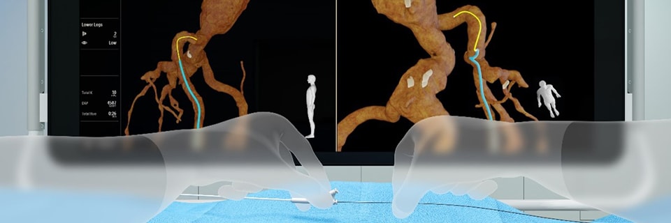
Fiber Optic RealShape (FORS) is a ground-breaking technology platform that is embedded into our LumiGuide solution. It enables real-time 3D visualization of the full shape of devices inside the body, without the need for fluoroscopy. FORS sends pulses of light through hair-thin optical fibers that run within minimally invasive devices. By sending laser light into the fiber and then analyzing how it is reflected back along the fiber, it is possible to reconstruct and visualize the full shape of the devices – in 3D, in real time, in distinctive colors and from any viewing angle. LumiGuide, powered by FORS, shows these intra-body devices in the context of the patient’s anatomy through integration with images obtained by conventional pre- or intra-operative techniques (CT, MRI and X-ray fluoroscopy respectively). Clinicians can therefore see more, perform fast while staying safe from radiation. LumiGuide is exclusively integrated within our Philips interventional X-ray systems. For more information on LumiGuide: www.philips.com/lumiguide For more information on FORS technology: www.philips.com/fors-technology
Innovation news and insights
You may also find the following articles interesting
Related pages
Related pages